
Esidrix
Discount 25mg esidrix free shipping
W ith atapem easure medicine look up drugs order esidrix online,fix theendatthehighest pointonthefundusandm easuretothesym physispubis. To avoidbias,dothiswith theblanksideof thetapefacing you,sothatyou onlyseethem easurem entonlifting thetape. Auscultation → U seanelectronic hand-heldD opplerfetalheartratem onitorasearlyas14weeks(F ig. Integrationlink:U terus inpregnancy Takenfrom R obbins& CotranPathologic Basisof D isease7E F igure7. Vaginalexam ination Inearly pregnancy A bim anualpelvic ex am inationshouldonlybeperform edif ultrasoundisnotavailabletoestablish gestation. U ltrasoundwillestablish gestationalage, confirm viability,ex cludeadnex alpathologyandshow m ultiplepregnancy. If avaginalex am inationhastobeperform edthesiz eof theuterusreflectsthegestation. Inlate pregnancy Vaginalex am inationallowsyou toassesscervicalstatusbeforeinductionof labour. F eelthedilatationandlength of thecervix,itsconsistencyand position,andthestation(level)of theheadaboveorbelow theischialspines(-or+respectivelyincm). Thecervix ism ostcom m onlylocated posteriorlyandtheheadof thefetusisfeltthrough theos. Antenatalinvestigations R outineandselectiveinvestigations Tim ing and/orindication Com m ent F ullbloodcount Booking,28weeks,36 Treatif haem oglobinlevelfallsbelow 100g/lwith ferrous weeks sulphate200m g t. R outineinm anyregionsof m ix edethnicity Bloodgrouping andantibodyscreen Booking andasadvised R hesusandK ellm ostcom m oncauseof isoim m uniz ation bylaboratory R ubella Booking (andafter contactif notim m une) H epatitisBandC Booking If hepatitisBantigenpositive,babywillrequirevaccinationsoon afterbirth. Bestto avoidbreastfeeding Syphilistesting Booking If positive,treatm entwith penicillinwillpreventcongenital syphilisinneonate U rinaryglucose E veryvisit W henpositive,reducesugarintake. If persists,carryout glucosetolerancetest U rinaryprotein E veryvisit Traceor+,checkm idstream urinespecim en. Confirm s gestationwithin10days U ltrasoundscanforplacentalsite Antepartum haem orrhage M orereliableasgestationadvanceswhenlowersegm ent after24weeks form s. If you donotcarryitoutyou m aym isspotentially seriousdiseaseandalsoleavesom em eninappropriatelyconcernedabouttheirperceivedgenitalsym ptom s. Aswellasrem em bering allthe principlesinChapter1you needtohavetheconfidencetoputpatientsattheireaseandtobeabletoreassurem enwith negativefindings,soex plore theirideas,concernsandex pectationswhentaking thehistory. A natom y Them alegenitaliaincludethepenis,scrotum,testes,epididym ides,sem inalvesiclesandtheprostategland(F ig. Allthestructuressupplying anddraining thetestes,including theneural,vascularandlym phatic supply,follow thisroute. Thisisclinically im portant,becausepainintherenaltractm aybereferredtothescrotum andtesticularcancerspreadstopara-aortic nodesratherthaninguinal nodes. Contractionof theejaculatoryductsandthe bladderneckcausesejaculationof sem en,followedbyincreasedtoneinthearteriolesanddiversionof bloodoutof thecorporaanddetum escence. Integrationlink:E rectionandejaculation-physiology Takenfrom Physiology5E Th e scrotum and its contents (F ig. Thescrotum isam uscularbag with thinpigm ented,rugoseskinandislinedbythedartosm uscle. Thisis highlycontractileandhelpstoregulatethetem peratureof thescrotalcontents,36°C being idealforsperm atogenesis. Along theposteriorborderof each testisistheepididym isform edby theefferenttubulesdraining thesem iniferoustubulesinthetestis. L ym phaticsfrom thepenisand scrotum draintotheregionalinguinalnodesthrough thesperm atic cord. Epididymitis ispainfulepididym alswelling andm aybeduetosex uallytransm ittedinfections,e. N eisseria gonorrh oeae orC h lamydia trach omatis,othercausesof non-gonococcalurethritis,ortourinarytractpathogenssuch asEsch erich ia coli. U reth ritis isinflam m ationof theurethraandm aycausedysuria oraureth raldisch arge. U nilateraltesticularatroph y m ayresultfrom m um psinfection,vascularcom prom iseafteringuinalherniarepairorfollowing alateorchidopex y foranundescendedtestis. A single testis m aybecausedbyanincom pletelydescendedtestisintheinguinalcanaloranectopic testisinthegroin,sochecktheseareas. Thism aycauserecurrentinfectionof theglanspenis (balanitis)orof theprepuce(posth itis)orboth (balanoposth itis). Them anm ayhaveproblem sfrom lossof libido,being unabletoachieveorm aintainan erection,prem atureejaculation,delayedejaculationorfailuretoejaculateandinabilitytoachieveorgasm. Alwaysclarifytheex actproblem and considercausesincluding psychological,alcohol,system ic disease(diabetesm ellitus)anddrugs;soyourhistoryneedstobewide-ranging. F indout whenthepainstartedandwhetheritisaccom paniedbyfeverandothersystem ic sym ptom s. Inepididym o-orchitisyou m ayfeeltheenlargedtenderepididym is separatelyintheearlystages,andthepainusuallycom esonm oregradually. How to takeaurethralswab M akesurethem anhasnotpassedurineforatleast2hours R etracttheprepuce U sing afinecottonbudorplastic loop takeasam pleof thedischargefrom theurethra L ookataGram -stainedsm earunderthem icroscopeim m ediately Platetheswab im m ediatelyonagarplates If thisisnotpossibleuseacharcoaltransportm edium Sam plesneedtoarrivewith thelaboratorywithin12hourstoensuredetectionof delicateorganism ssuch asN. Checkthegroin, perineum andscrotalskinfortheserashesandintertrigo(infectedecz em a)intheskincreasesandecz em a (anothernam eforderm atitis). Thepenileskinhasm oresebaceousfolliclesthanm ostotherareas;enlargedfolliclesare som etim esm istakenforwarts. N um erousuniform pearlypenilepapulesaroundthecoronaof theglansarea com m onnorm alfinding. Askthem anwhetherthisisrecentandif ithascausedproblem ssuch asinfection,spraying during m icturitionorsex ualdifficulty. R em em bertodraw theforeskinforwardafter ex am inationtoavoidparaph imosis,which ispainfuloedem aof theglans. Takeaurethralswab if yourpatient hasadischargeorishaving sex ualhealth screening (Table7. If eitheryou orthepatientiscold,thecrem asterm usclewillcontractandyou willnotbeableto palpatethetestesproperly. F eeltheanteriorsurfaceandm edialborderwith yourthum b andthelateralborderwith yourindex finger. Verygentlysqueez ethe fingersof yourlefthandtopush thetestisinferiorlyandallow you toassesstheupperpoleof theorgan. U sethisopportunitytoencourageallyounger m entoex am inetheirtesticlesregularly-testicularself-ex am ination. You willbeableto feelthesperm atic cordandwithinitthevasdeferenswhich feelslikeathickpieceof string (F ig. Inadarkenedroom holdapen-torch againstthe swelling andseeif lightistransm ittedasabrightredglow totheskinof thescrotum. Thisconfirm sitis fluid-filled,whereasnotransm issionof lightoccurswith asolidm ass. Anogenitalregion Theanalm argin,rectum andprostateglandshouldallbeex am inedaspartof them alegenitalassessm ent. Perianalwartsarecom m on,oftenin associationwith genitalwarts(condylom ataacum inata). Integrationlink:Prostatic cancer-pathology Takenfrom R obbins& CotranPathologic Basisof D isease7E U ltrasound ♦ K eypoints page221 page222 ♦ D onotavoidex am inationof them alegenitaliasim plybecausethepatientsaysitisallnorm al. U ltrasoundex am inationof thescrotalcontentsishelpfulindifferentiating solidfrom cystic lesions. If you cannotpalpateanyabnorm alitythenthereis nopointinperform ing anultrasoundex am ination,itwillnotrevealanym oredetails. Theupperendof theanalcanalis definedbythepuborectalism uscle,which contractsif thepatientcoughsorconsciouslysqueez esthepelvic m uscles. W henthepatientislying intheleftlateralpositiontherectum passesbackwardsandupwardsfollowing thecurveof coccyx andsacrum posteriorly. Anteriorly,inascending order,liethem em branousurethra,theprostateandthebladderinm en;andthevaginaandcervix inwom en(F ig. The upperandanteriortwo-thirdsof therectum iscoveredinperitoneum reflectedontothebladderbaseinm enandthepouch of D ouglasinwom en. N orm allyitfeels rubberyandsm ooth andhasam ediangrooveseparating sym m etricalleftandrightlobes. Theonlypalpablem assesshouldbeex ternaltotherectum andnorm allyaretheprostateinm enandthecervix inwom en.

Buy esidrix 25 mg amex
In such patients symptoms magnesium deficiency order 12.5 mg esidrix with visa, the severely damaged cause an increase in serum phosphorus concentra kidneys cannot respond to further increments in tion. Also, the concentra calcium and phosphorus from the skeleton into tion of serum phosphorus may fall during refeed the extracellular fluid. This phosphorus cannot ing after a period of calorie or protein malnutri be excreted by the kidney and hence serum tion. The same phe the use of calcium compounds in patients nomenon occurs in dialysis patients. First, the in the serum levels of phosphorus due to the levels of serum calcium and phosphorus are ability of these compounds to bind phosphate in higher in patients with advanced kidney failure the intestine. Second, following total or subtotal be followed by a reduction in serum levels of parathyroidectomy in patients with kidney fail serum phosphorus as discussed above. As kidney function deterio phosphorus levels not only may remain above rates further, an absolute vitamin D-deficient normal but may rebound rapidly after dialysis to state develops, with the blood levels of predialysis levels. Multiple reasons may account for these disturbances in the function of its target organs: variations (Table 5). The 2 major types of bone parathyroid glands, bone, intestine, and skeletal disease that are commonly encountered in pa muscle (Table 4). In this tive or absolute deficiency of vitamin D or its condition, there is also increased bone formation active metabolites and/or resistance to their ac as evidenced by increased amounts of osteoid. This osteitis fibrosa is a high-turnover bone dis Vitamin D may affect mineralization through ease. It is not evident whether a fective mineralization of osteoid leads to rickets deficiency in one or more of the vitamin D in children and osteomalacia in adults. Osteomalacia may be more frequently excess osteoid does not necessarily mean osteo encountered in uremic patients with low blood malacia. These derangements result in a cline labeling can differentiate between these 2 defect in collagen cross-linking and may affect possibilities and is thus critical for the diagnosis bone mineralization. The skeleton in osteomalacia is lagen metabolism are most likely due to vitamin weakened, and patients with this bone disease Ddeficiency. The magnesium content of the the most important factor in the development of bones of these patients is increased, and this may osteomalacia is aluminum overload. This type of bone cium intake is increased or if dialysate calcium is disease has been called low-turnover bone dis high. Osteoscle seen in dialysis patients who have a large content rosis appears as increased bone density in roent of aluminum in bone and in whom the aluminum genographic studies. Histologically, osteosclero is localized in the mineralization front (ie, the sis is most likely due to accumulation of limit between osteoid and calcified tissue). With unmineralized trabecular bone with an increase a decrease in the use of aluminum-containing in total bone mass. Because osteosclerosis af compounds for the control of hyperphosphatemia, fects trabecular bone, it is most evident in the the incidence and prevalence of osteomalacia vertebrae, pelvis, ribs, clavicles, and metaphyses have been decreasing. Increased burden of iron, of long bones, which are made predominantly of alone or in combination with aluminum, can cancellous (trabecular) bone. Certain experimental and clini failure patients before and after treatment with cal evidence suggests that osteosclerosis could peritoneal dialysis or hemodialysis. Immobiliza characterized by a defect in bone matrix forma tion, calcium deficiency per se, and chronic pro tion and mineralization, increased osteoid thick tein depletion may be causes of the osteoporotic ness, and a decrease in the number of both component of kidney osteodystrophy. There older than 50 years, factors that cause postmeno are no excessive amounts of aluminum in the pausal, idiopathic, or senile osteoporosis may mineralization front. Certain factor(s) that may act locally als once the labile calcium carbonate component to inhibit calcification and are present in the of bone is lost. Although there may be a slight blood of these patients may possibly be removed improvement in negative calcium balance follow during hemodialysis. Local tissue injury may ing treatment of the chronic acidosis with alkali, also predispose to calcification when the calcium a positive balance for calcium usually does not phosphorus product is normal or only slightly occur, and hypocalcemia, bone pain, and radio elevated. More certain proteins involved in prevention of calcifi over, there is no convincing evidence suggesting cation has been demonstrated in macrophages that chronic acidosis can cause defective mineral and smooth muscle cells of blood vessel walls. An increase in the calcium-phosphorus vascular calcification seen in patients with kid product in the extracellular fluid is probably the ney failure. The inci the chemical nature of soft-tissue calcifica dence of soft-tissue calcification is high when the tion may vary in different tissues. Thus, the calcium-phosphorus product (each in mg/dL) calcification found in nonvisceral tissue (periar exceeds 70, while soft-tissue calcification is infre ticular and vascular calcification) consists of quently noted when the calcium-phosphorus prod hydroxyapatite, with a molar Ca:Mg:P ratio simi uct is below 50. These breakpoints not withstand ing, and because of the biological variations in lar to that of bone. These observations suggest that the mechanisms Alkalemia, which often occurs after hemodialy responsible for the calcification of various tis sis, may persist during the interdialytic period sues in uremic patients may be different. The calcifica tion in the eye may produce visible inflammation tion appears as a fine, granular density outlining and local irritation, resulting in the red eye of a portion of the entire artery, giving a radio uremia. This is a transient phenomenon and may graphic appearance of a pipestem due to deposi last only a few days. Recurrence of the red eye tion of calcium within the media and the internal phenomenon is not infrequent, and it becomes elastic membrane of the artery. The lumen of the apparent each time a new calcium deposition vessel is usually not involved. More commonly, con fication may first be seen in the dorsalis pedis as junctival calcium deposits are asymptomatic and a ring or a tube as it descends between the first are seen as white plaques or as small punctate and second metatarsals. Calcification can also deposits on the lateral or medial segment of the occur in atherosclerotic plaques in the intima of bulbar conjunctiva. Also, calcium deposits may large vessels whose radiographic appearance is occur within the cornea at the lateral or medial that of discrete, irregular densities. It is possible segments of the limbus, the so-called band kera that uremic patients are more prone to this type topathy. Slit-lamp examination permits easier of calcification because of the presence of hyper recognition of these lesions. Vascular calcifica may be found in the lungs, stomach, myocar tions are seen in kidney failure patients and in dium, skeletal muscles, and kidney. These calci those treated with hemodialysis, and they persist fication are usually not evident radiographically, after kidney transplantation. Congestive heart failure, car reported incidence of arterial calcification in diac arrhythmias, and heart block may occur in creases with duration of dialysis treatment. In a patients with calcium deposition in the myocar series of 135 patients published in 1977, the dium or in and around the conduction system of incidence of vascular calcification increased from the heart or the mitral annulus. Calcification of 27% in those treated for less than 1 year to 83% cardiac valves are not infrequent. Such patients may have ery artery and has been seen in arteries of the reduced vital capacity and reduced carbon mon forearm, wrist, hands, eyes, feet, abdominal cav oxide diffusion. The calcification tion has been noted after subtotal parathyroidec may be very extensive, rendering the artery so tomy in these patients. Extensive pulmonary rigid that the pulse is not palpable and the Korot calcification may lead to severe pulmonary fibro koff sounds may be difficult to hear during the sis, pulmonary hypertension, and right ventricu measurement of the blood pressure. Calcification of the heart and cation may also present difficulties during sur lung constitute a major risk factor for increased gery for the creation of arteriovenous shunts or morbidity and mortality in dialysis patients. This may be associated regress; in some patients, improvement or disap with marked deposition of calcium oxalate in pearance of arterial calcification occurs within soft tissues. Such deposition in the myocardium, months to years after subtotal parathyroidectomy or mitral and aortic valves, can cause cardiomy or renal transplantation. The inci drome characterized by the development of pro dence of periarticular calcification varies widely gressive ischemic skin ulcerations involving the among dialysis patients. These calcification were fingers, toes, thighs, legs, and ankles has been absent in 1 report but were encountered in up to observed in a small number of patients with 52% of the patients in other series of dialysis advanced kidney failure. The incidence of periarticular calcifica in patients after successful kidney transplanta tion may increase with the duration of dialysis. With better common among patients treated with continuous control of serum levels of phosphorus, this type ambulatory peritoneal dialysis. The patients almost always have vascular cal Periarticular calcification may be detected be cification involving the media of the arteries, and cause of the pain induced by the deposition of they usually exhibit X-ray evidence of subperios calcium or may be noted by routine X-ray exami teal bone resorption. A period of as small discrete radiodensities around the shoul hyperphosphatemia has been present for some ders, wrists, phalangeal joints, hips, or ankles. The Tendosynovitis or tendonitis with abrupt pain lesions may be preceded or accompanied by may develop, presumably caused by deposition severe pain. The synovial or tissue necrosis, tender, slightly erythematous, fluid of the involved joints is clear with normal subcutaneous nodules may develop, or there may viscosity and number of cells.
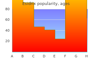
| Comparative prices of Esidrix | ||
| # | Retailer | Average price |
| 1 | True Value | 282 |
| 2 | Ace Hardware | 608 |
| 3 | Dillard's | 948 |
| 4 | ShopRite | 823 |
| 5 | Wal-Mart | 264 |
| 6 | Sears Holdings | 331 |
| 7 | Burlington Coat Factory | 670 |
| 8 | Albertsons | 448 |
Buy generic esidrix 25mg on line
Hum mol Genet 2006;15:1049 elements symptoms 5 dpo discount 12.5mg esidrix, common dietary sources of these elements and 58. Increased serum levels of a parathyroid hormone-like protein in malignancy-associated hypercalcemia. Studies of the mechanism by which phosphate infusion lowers serum calcium concentration. We intend to draft a practical guideline, focusing on operationalized recommendations deemed to be useful in the daily management of patients. This systematic search found 1100 articles, which was reduced to 312 based on title and abstract. The working group assessed these for eligibility in more detail,and32full-textarticleswereassessed. Data on quality of life and the risk of complications have just started to emerge, and clinical trials on how to optimize therapy are essentially non-existent. This guideline is therefore mainly based on how patients are managed in clinical practice, as reported in small case series and based on the experiences of the authors. See further We formally graded only the evidence underlying rec section ‘Summary of methods used for guideline ommendations for therapeutic choices and target calcium development’. Monitoring of the patient when implementing different therapeutic efforts, aiming to achieve the therapeutic goals. Predisposing factors involved are syndrome (7)) or as an isolated endocrinopathy young age, female gender, Graves’ disease, lymphadenect (see Table 1). Impaired renal function has been associated secretion immediately after surgery resulting in hypocal with the age of the patient, duration of the disease and caemia (14, 15). Normal to high need to mineralize the foetal skeleton and to provide this calcium levels at 1 month after thyroidectomy appears to essential mineral to the breast milk to sustain postnatal increase the chance of parathyroid gland recovery. The skeletal development and well-being in the newborn (25, clinical and disease-related variables do not seem to 26, 27, 28, 29). In effects on the kidney prompt urinary calcium levels to fall support of these issues, large cohort studies from Denmark during lactation. Also suggested are 24-h urinary calcium excretion feeding and hypotonia) may result (27, 29, 30, 31, 32, 33, to be measured yearly or every other year. Conversely, maternal overtreatment resulting in hypercalcaemia can suppress foetal parathyroid 2. In recent years, a number of studies have been published showing that normocalcaemia can be main 2. The treatment should aim to maintain serum calcium to its continuous episodic endogenous pattern of levels in the low normal range, serum phosphate within the secretion. Hyperphosphatae ations in serum calcium levels compared with injections mia should be addressed by decreasing dietary phosphate once-a-day (43). Magnesium-depleted patients should calcium significantly compared with twice-a-day injection be replete with this mineral. It is interest in calcium metabolism might also find the unknown whether long-term therapy also may cause guideline useful, as might our patients. According to a predefined schedule, daily Congress of Endocrinology, 18th May 2015 in Dublin. Aims a daily dose of 25 mg or up-titrated to a daily injection of the overall purpose of this guideline is to provide either 75 or 100 mg. Guideline working group systematic approach for synthesizing evidence and grad this guideline was developed and solely sponsored by the ing of recommendations (45). The other members: C Marcocci, the first methodological step for the guideline develop L Rejnmark and D M Shoback (endocrinologists), W van ment group is to define the clinical question(s) to be Biesen (nephrologist) and A Sitges-Serra (endocrine addressed in the guideline (see section ‘Clinical question, surgeon) were all approved by the Committee. These clinical working group had two in-person meetings (January and questionsareformulatedinsuchwaysthattheiranswerscan December 2014) and communicated by phone and email. Prior to the process, all participants Whereas the question, is hydrochlorothiazide better than completed conflict of interest forms. Importantly, the clinical is judged as not important or if a management strategy questions should be specific in terms of populations. Another available evidence, the guideline development group rated consideration is that you cannot abstain from recommen clinical outcomes either as critical, important but non dations when there is no evidence, as treatment decisions critical, and not important (46), see section ‘Clinical need to be made any way. To optimize transparency, all recommen relevant articles, synthesis of the evidence was made in dations provided are accompanied by text explaining why two directions: i) estimation of an average effect for specific recommendations were made. Clinical question, eligibility criteria and endpoint is provided as a summary rating (using four categories: definition high, moderate, low and very low) across studies for each outcome, as it may be that quality differs for different the clinical question on which the literature search was outcomes. The recommendations are worded as recommend (strong recommendation) and suggest (weak the following outcomes were rated by the guideline recommendation). The meaning of a strong recommen working group as critical: dation can be stated as follows: reasonably informed i) Mortality. Only for recommen defined as symptoms or episodes of nephrolithiasis dations regarding treatment, did we aim for formal or nephrocalcinosis. Only quality evidence may not directly translate into a strong articles in Dutch, English, German and French were Sample sizes were too small and follow-up too short well as the relevant interventions and outcome measures. Articles identified by the working group as relevant were used to optimize the search strategy. Articles Cramps, tetany and seizures " Occurrence of cramps, found by ‘snowballing’ were also considered. For rence of these conditions under different treatment the final recommendations, other literature (basic litera regimens. Conclusions from scientific literature and reported in various ways on serum calcium levels. In directions for further research general, calcium levels within the target range can be achieved with different treatment (combinations), and the 4. Summary and interpretation of the main findings risk of severe hypo or hypercalcaemia is low. Study results in perspective definition (see section ‘Clinical question, eligibility Very little evidence is available on how best to treat criteria and endpoint definition’). Data on QoL and the risk of complications have had a sample size of O100 patients. Also no studies are available that relate target calcium levels to clinically relevant endpoints. Study endpoints Thus, we do not know what the optimal calcium target Mortality " Only one study reported data on mortality is. No deaths were reported in this randomized study to formulate recommendations based on strict evidence. If the patient’s well being is improved by titrating treatment in such a manner Reasoning " Hypocalcaemia is defined as an ionized or that serum calcium levels are in the upper part of the albumin adjusted serum total calcium level below the reference interval, this may be accepted as no data exist on lower limit of the reference interval. Measurements of whether relatively high normal serum calcium levels are of ionized or total calcium mostly reflect local traditions, specific harm to patients. It is generally assumed that this is exists on long-term benefits (or harms) of achieving these related to high serum phosphate levels and an increased therapeutic goals. Accordingly, patients cannot be ensured serum calcium–phosphate product, which is why it seems long-term beneficial effects, when accepting potential side reasonable to aim at keeping phosphate levels within the effects to drug treatment or (troublesome) changes in normal range (59). Thus, we consider it inappropriate management, if therapeutic efforts have immediate R. Moreover, low magnesium levels may monitoring of patients at regular time intervals. Accordingly, calcium levels may, however, change and complications it seems reasonable to aim at keeping serum magnesium may emerge at any time, with or without any apparent levels within the reference range. Thus, we find it of importance to empower patients with knowledge of symptoms and co-morbidities and drugs R. If a patient is diagnosed with one of the have neuromuscular complaints (4, 38, 53). To ensure that diseases or initiates treatment with one of the drugs, this symptoms are not caused by low vitamin D levels, it seems may necessitate changes in the medical treatment of reasonable to ensure an adequate vitamin D status (55).

Buy esidrix online pills
By measuring the diameter of the lesion (assuming that it is approximately spherical) and calculating its volume with the formula: volume ¼ 4=3pr3 where p is the constant pi and r is the radius of the lesion treatment yeast diaper rash generic 12.5mg esidrix amex. After the volume is calculated on two separate occasions, doubling time can be derived from a plot of volume versus time. A calculation that describes the effect of size and other factors on slowing of tumor growth. Doubling time calculation is a rough estimate because it assumes simple growth kinetics and the absence of other factors that affect tumor growth. However, tumor cell populations exhibit a reduction in growth rate with increasing size because they receive less blood supply to the center of the tumor as the mass grows, and the Gompertz equation accounts for this effect. The vast majority of infections originate from the patient’s own endogenous flora. In investigating the cause of an infection, cultures should include blood, urine, sputum, and if appropriate to the patient’s clinical status, stool, pleural fluid, or peritoneal fluid. Renal cell carcinoma and multiple myeloma tend to be purely lytic, prostate carcinoma tends to be mostly blastic, and other bone lesions are mixed. Lytic bone lesions are often associated with hypercalcemia, unlike blastic metastases. A dull, aching discomfort that is worse at night and may improve with physical activity. Most types of tumors can metastasize to the lungs; therefore, the more common the tumor, the more common the lung metastases. Tumors that spread via the bloodstream, such as sarcomas, renal cell carcinoma, and colon cancer, tend to produce nodular lung lesions. Those that spread via lymphatic routes, such as cancers of the breast, lung, pancreas, stomach, and liver, may manifest a pattern of lymphangitic spread. Headache occurs in up to 50% of patients with intracranial metastases and is classically described as occurring early in the morning, disappearing or decreasing after arising, and associated with nausea and/or projectile vomiting. Other symptoms include focal signs such as unilateral weakness, numbness, seizures, or cranial nerve abnormalities. By decreasing intracranial pressure with steroids, followed by definitive therapy. Surgery is recommended for patients with single intracranial lesions if technically possible, whereas radiation therapy is generally administered for multiple lesions. Chemotherapy may also be used, but the results are not as reliable as the other modalities owing to the difficulty of chemotherapy agents penetrating the blood-brain barrier. Frequently similar to the symptoms of heart failure with dyspnea, peripheral edema, and an enlarged heart on chest x-ray. However, the dyspnea is often out of proportion to the degree of pulmonary congestion seen on the x-ray. Kussmaul’s sign, or jugulovenous distention with inspiration, and pulsus paradoxus of > 10 mmHg with distant heart sounds are clues to the presence of a pericardial effusion. Treatment depends on the patient’s condition but should include drainage of the fluid for diagnostic as well as therapeutic reasons. A nonsurgical approach is preferred, with catheter drainage followed by sclerosis of the pericardium, sometimes with a sclerosing agent such as doxycycline. Other approaches include subxiphoid pericardiectomy, balloon pericardiectomy, pericardial window, and pericardial stripping for patients with prolonged life expectancy. Other symptoms include lower extremity weakness, bowel or bladder incontinence, or increased deep tendon reflexes in the lower extremities. Once neurologic symptoms appear, the nerve damage may be irreversible; therefore, early diagnosis of cord compression is essential. Initially by decreasing spinal cord swelling and pain with high-dose steroids and adequate pain medication. Definitive treatment with surgery or radiation therapy must be carried out emergently to prevent irreversible neurologic deterioration. This paraneoplastic syndrome is also known as “marantic endocarditis” and has also been described in other types of cancers. Usually the appearance of embolic peripheral or cerebral vascular events causing arterial insufficiency, encephalopathy, or focal neurologic defects. The emboli originate from sterile, verrucous, fibrin-platelet vegetations that accumulate on the heart valves, likely due to a hypercoagulable state from malignancy. However, echocardiograms may be negative, and the diagnosis is usually made postmortem. Treatment with anticoagulants or antiplatelet drugs has been tried with little success. Electrolyte and metabolic disturbances such as hyperuricemia, hyperkalemia, hyperphosphatemia, and hypocalcemia that can result in renal failure, arrhythmias, and seizures. These disturbances occur when rapidly growing tumors are effectively treated with chemotherapy and breakdown products of dying tumor cells are released in large amounts into the bloodstream. The complication is seen within hours to days after treatment of malignancies such as acute leukemia and high-grade lymphomas such as Burkitt’s lymphoma. With allopurinol and supportive measures for renal failure such as vigorous hydration, dialysis if necessary, and appropriate treatment of electrolyte disorders. Rasburicase (recombinant urate oxidase) can be administered when uric acid levels are not lowered by standard approaches. Prophylactic treatment with aggressive hydration and allopurinol can prevent this serious complication and should always be given before chemotherapy in malignancies with high proliferative index. Pain medications are to be administered in a stepped approach according to the intensity and pathophysiology of symptoms and individual requirements. Patients with moderate-to-severe pain generally require an opioid agent such as codeine or oxycodone; severe pain requires a stronger opioid such as morphine. Neurol Clin 16:449–481, 1998; and Didelot A, Honnorat J: Update on paraneoplastic neurological syndromes. The incidence is increased and nearly equal in men and women, in China, Africa, Russia, Japan, Scotland, and the Caspian region of Iran. In the United States, African American men living in urban areas are at increased risk. Adenocarcinoma of the esophagus is now more prevalent than squamous cell carcinoma in the United States and Western Europe, with most tumors located in the distal esophagus and esophagogastric junction. Fewer than half of patients appear to be operable at the time of presentation, and of these, only one half to two thirds have tumors that are completely resectable. Nonsurgical patients are treated with combined chemoradiotherapy or palliative measures if their performance status is too poor for active therapy. Some evidence indicates that survival in patients with adenocarcinoma of the esophagus is improved with preoperative combined chemotherapy and radiotherapy. Ongoing trials are investigating whether the outcome with chemoradiotherapy is equivalent to that of surgery. Differences between mutations associated with the intestinal and diffuse types of gastric cancers may account for their different natural histories. Describe the diagnostic and staging evaluation for patients suspected of having pancreatic cancer. How is the diagnosis of pancreatic cancer confirmed and the extent of metastatic disease evaluated? Additional staging includes routine laboratory studies, chest x-ray, and other tests as directed by the history and physical. Which environmental factors are thought to be related to the development of colon cancer? A syndrome characterized by the occurrence of thousands of adenomatous polyps throughout the large bowel. If left untreated, cancer will develop in all patients with this syndrome, usually before the age of 40. The most common hereditary colon cancer syndrome that is also associated with extracolonic cancers such as endometrial, ovarian, pancreatic, gastric, renal, hepatic, and small bowel. If elevated, it should be retested 30–45 days after complete resection of the cancer.
Order esidrix 25mg overnight delivery
Poultry: There are several methods of slaughter: wringing the neck treatment with cold medical term buy esidrix online pills, dislocating the neck or beheading. The following guidelines are for any species that may be consumed in the field environment. General: Condemn animals with gross contamination of interior surfaces or organ systems and/or discoloration of peritoneal or pleural cavities. Generalized abscesses, emaciation, and jaundiced organs or tissues are reasons for condemnation. Lymphadenopathy indicates disease or inflammation in the area drained by the enlarged nodes. Local adenopathy may indicate a local process only (condemn only affected area), while more extensive adenopathy probably implies widespread disease process. Palpate and examine lymph nodes of the head and neck for gross swellings or lesions. Inspect and palpate all surfaces for abnormalities, discoloration, masses and parasites; examine the heart, lungs and diaphragm as well. Slice open organs and examine for parasites, infection or disease states such as tumors. Joints and Skeletal Muscles: Bruises and localized lesions may be removed and the rest of the carcass consumed. For arthritic and swollen joints, remove affected limb, and then consume carcass if arthritis is not due to systemic disease such as septicemia or caseous (cheese-like) lymphadenitis. Neoplasia, Tumors or Abnormal Growths: Condemn organ system and/or carcass if spread throughout. Off Odors: Condemn carcass with strong odors of urine, ketones (a fruity smell) or pungent sexual odors. When excess food must be stored and preserved for future use, follow these rules: preserve and store only wholesome foods that were initially safe to eat; use only potable water and spices when curing or preserving food; cold storage/freezing is the best method if available; periodic re-examination of stored products is essential to ensure wholesomeness and prevent consumption of contaminated or deteriorated food (moldy, infested, stale). What You Need: Knife, meat, potable water, salt, 1% salt solution (brine), string, green hardwoods, building, saltpeter (potassium nitrate), spices, fire source, hay, salt box and/or brine pan, boiling pot. Curing Although it may be done alone, curing should be done in association with smoking. Raw meat should be clean, edible and sliced against the grain into manageable pieces (step one of beef prep for smoking). If using brine, then the solution should be 1% salt (one pound of salt to 9 pints water). Use clean plastic, glass or earthenware containers, not wood or metal containers to hold brine solutions. Smoking There are several acceptable procedures for smoking meat and different step by step processes. Smokehouse Use any well-sealed building with a vented roof and a floor that can have a fire pit. Let fire burn down to coals and then stoke it with green wood to produce “cold smoke” (less than 85°F). Allow even smoking and avoid contact spoilage by ensuring that all meat hangs free. Time Smoke meat for 4-5 days, depending on size of house, size and number of pieces of meat to smoke. The hole for string should be centralized enough to prevent meat ripping during smoking. Hang meat and prepare smoking record (see preservation records and recommendations below). Smoked meat should be edible for up to one year depending on climate, condition of meat prior to smoking and insect and rodent control. Meat may also be dried over slow coals or sun-dried (sprinkle with pepper and hang about 20 ft into air above insect line). Pemmican Two basic ingredients: lean meat that is not salt cured and rendered fat. Prepare a casing, such as intestine, by cleaning (strip out contents and boil) and tying one end. The fat will separate into tallow, the liquefied oil from fat, and (cracklings), the fat residue. For pickling, mix 50 pounds of salt and 5 pounds of saltpeter with 20 gallons of water. Canning and Other Methods these procedures are effective but require resources and equipment not readily available in a field environment. Records should have the following information at a minimum: meat type, date, source of meat, weight and cut of meat, total time cured (preserved), wood used and/or type and amount of salt/seasoning/brine used. What Not To Do: Do not use meat that is unfit for consumption based on ante or postmortem exams. What You Need: Rope, twitch, nose lead, stethoscope, pen, paper, leather gloves, exam gloves, light source, rectal thermometer (large animal style preferred) What To Do: 1. Restraint: Allow owner and/or indigenous persons to handle and restrain the animals as much as possible. This is probably the most difficult part of the examination and may be the most dangerous. Halter Fasten a rope loop around the animal’s neck with a bowline knot to make a temporary rope halter. Pull a bight of the standing end through the loop from rear to front and place over the animal’s nose. A twitch is a small loop of rope or smooth chain twisted around the upper lip of the horse to divert attention from work being done elsewhere on the horse. Twist the rope or chain with a stick or rod to tighten the twitch, but avoid circulatory compromise. Casting a cow (Burley Method) You will need approximately 40 ft of rope, with the center of the rope over the withers and wrapped as shown in the diagram. While maintaining control of the head, pull tightly on the ends of the rope and the cow will fall. To tie the rear legs, keep both ropes taut and slide the uppermost rope along the undersurface of the rear leg to the fetlock. Then carry the end around the leg and above the hock, across the cannon bone and back around the fetlock. Restraining the Legs Tie all four feet together to restrain the animal after it has been cast (dropped). Tie the other legs to this one alternately, first a front leg, then a 5-129 5-130 rear one and repeat. Cattle Tail Restraint Bend the tail of the cow toward the side or back of the animal to distract the cow. Examination: Once an animal is sufficiently and securely restrained, begin the physical exam. Remember: the diseases and injuries of animals can be similar to those in humans, but seek advice from appropriate veterinary providers or the Merck Veterinary Manual if available. One can only diagnose and treat based on his level of knowledge and understanding of veterinary medicine. Many zoonoses are threats in the field environment and precautions need to be taken to minimize them. Review the Preventive Medicine chapter and individual infectious disease sections for specifics on zoonoses and how to prevent them. Abuse is unethical, unnecessary and may jeopardize the relationship with native personnel. When to intervene is dependent on the state of parturition, the presentation of the fetus, duration of labor and history of underlying disease processes. This outline will provide only the basics of “normal” parturition and guidelines for observation and minimal intervention. Subjective: Symptoms Prior to parturition a normal animal will walk with difficulty, often looking back at her flanks. The udder may swell and become distended with milk, the tailhead ligament will relax and the vulva may swell and begin to discharge mucus or fluid. Duration of labor varies considerably between species (15 minutes for horse; up to 7 hours for a pig litter), and is longer in animals giving birth for the first time. He/she may also be of assistance in controlling the animal and giving medical history. Animal will present with an enlarged abdomen and a drop in body temperature (1-2° below normal 12-24 hours before birth–see Physical Exam section of this chapter).
Syndromes
- Permanent skin damage and scarring (very rare)
- Choking (coughing, shortness of breath)
- Fatigue
- Eye discomfort
- Abnormal placement of the heart toward the right side of the chest instead of the left
- Cardiomyopathy with heart failure

Order esidrix online now
T em poral Th e locationh alfway upth e nail Suggests illness 3 month s ago relationsh ips (location ofth e line tells w h en th e illness w as experienced) N ote th e 2 Beau’s lines A bout2 month s apart T h in B rittle N ails symptoms 7dpo generic 12.5mg esidrix with amex. S evere m alnutrition N ote th e th innails inth is wom an with severe osteopenia S ystem ic A m yloidosis C entralN ailR idge. C auses – Irondeficiency – F olicacid deficiency – P roteindeficiency C entralN ailC anal (M edian N ailDystroph y). A ssociations – S evere arterial disease – S evere m alnutrition – R epetitive traum a N ailP itting. C onditions – P soriasis (random appearance ofpits) – A lopecia areata (geom etricrippled grid) – Ecz em a – L ich enplanus Images courtesy of Courtesy of Sarah Caney < in some situations (eg, unexplained However, in cats its diagnostic value renomegaly) a kidney biopsy or fine appears compromised by overlap in needle aspiration may be desirable. This is valuable as the which cats will develop azotaemia has stage (severity) of disease is related to not been demonstrated. Courtesy of Jessica Quimby and can help to focus attention on Other assays appropriate treatments. Full history Evaluation of progress, complications and term, even if owner concerns Following diagnosis (and stabilisa Evaluation of changes since last assessment stable, cats tion, if necessary), initial re-evaluations Full clinical Body weight, % change in body weight and body should typically be undertaken every examination condition score should be 1–4 weeks, according to clinical needs. In the long term, even if stable, cats including thyroxine, liver enzymes and acid–base should be re-evaluated at least every 3–6 months. Particular status attention should be paid to appropriate monitoring of the effica Diagnostic Ultrasonography or radiography (Figures 6 and cy of interventions to ensure that therapeutic targets are being imaging 7) – to assess for structural changes, obstructions met. In advanced disease, care may be needed to avoid exacer or other lesions – should be part of the initial investigation and may be worth repeating, bating anaemia by too frequent blood collection. This will facilitate individualised tion, fluid therapy may be beneficial in management plans to be created that take into addressing electrolyte and acid–base distur consideration the wishes and ability of the bances, and diluting uraemic toxins. Importantly, palatable and easy to administer taemia is stable, fluids should be tapered over medications make this easier, and it may be possible to combine medications 2–3 days before the patient is discharged. The patient should be thoroughly re-evaluated each time significant diet where possible is important, as it will progression has occurred to ensure treatable complications are not being 48 also increase water intake. Courtesy of Sarah Caney Figure 8 Multiple sources of fresh water are important for maintaining hydration. Courtesy of Jessica Quimby dehydration is a concern, but it is important to ensure intake of other nutrients is maintained. A feeding tube is suitable for long-term maintenance of hydration and is Renal diets a more physiological approach. Careful attention to body condition, muscle mass shown to and caloric intake is important. Courtesy of Jessica Quimby subcutaneous fluid therapy (75–150 ml every 1–3 days) can be used on an outpatient basis significantly (Figure 9) or by owners at home to maintain Protein and phosphate restriction hydration. Although a balanced electrolyte solution Protein restriction and phosphate restriction such as lactated Ringer’s solution is often used, are considered together as they are the main a hypotonic solution (half-strength lactated features of commercial renal diets, and are Ringer’s or 0. Fluids can be administered via a protein per 100 kcal (above the 5 g/100 kcal needle and giving set, or through an indwelling recommended allowance for adult cats,51 but subcutaneous catheter, although there is the below the 9–10 g/100 kcal commonly seen in risk of the latter becoming blocked or infected. Renal formulated Studies evaluating effect restriction, renal diets contain Table 3 diets are restricted in both protein of renal diets on longevity much less phosphate compared and phosphorus, but other with typical maintenance diets. Where diet alone is insufficient, the use of Appropriate home-prepared diets64 (see box intestinal phosphate binders is important. Senior diets generally have lower protein (and phosphate) than adult maintenance diets but, in the absence of a renal diet, earlier intervention with a phosphate binder may be necessary (see Table 4). Additionally, formu renal diet cannot be used, or is insufficient to lations of calcitriol can make accurate dosing control serum phosphate, phosphate binders difficult in cats. Hyperphosphataemia should should be used (given with food), the response also be carefully controlled when using this monitored (eg, 1 month after medication therapy to avoid increasing the serum change), and the dose adjusted accordingly. Although not subjected to clinical testing, the Panel suggests adopting the target serum phos phate concentrations recommended by iRiS:71 Panel recommendations: calcitriol < Stage 2 disease: 0. Supplementation with Quality of evidence as an intervention potassium gluconate (or citrate) is recommend < Increased longevity: No evidence of benefit ed if serum K <3. However, even when cats are calm and a standardised protocol is followed,105 Other nutrients measurements will vary with the equipment, the operator, the cat and the circum stances. Clinical efficacy as antihypertensive uncertain and best currently considered as adjuvant therapy in refractory cases Benazepril 0. Diagnosis is supported by a bone marrow aspirate/core and cats become transfusion dependent for months. The target for therapy should be a 37,42,44,120,123 known to carry a poorer prognosis. Very similar < oral iron supplements (ferrous sulfate): 50–100 mg/cat per 120 findings have also been reported elsewhere. This procedure has numerous implications including ethical, Anabolic steroids financial, welfare and monitoring considera information regarding the efficacy of anabolic tions. Their main demonstrated beneficial effects; and with indications are for management of acute kidney some techniques adverse effects occur. Decisions on therapy should be made on a case is also frequently associated with significantly elevated parathyroid by-case basis, assessing risks and benefits. There is, however, hormone concentrations that may potentially complicate existing evidence that low dose (0. Close monitoring of routes (eg, benazepril,182 telmisartan183) are preferred where possi the cat’s clinical condition, serum creatinine and thyroxine is ble. Nevertheless, the risk:benefit ratio of each treatment should be required to tailor the dose for each patient. Starting doses can be assessed,184 and dose adjustments may help to mitigate risks. Case-control disorders recorded in cats attending primary-care veteri study of risk factors associated with feline and canine nary practices in England. Risk factors associat chronic renal disease in Australian cats: a prospective study ed with the development of chronic kidney disease in cats of 184 cases. Diet and lifestyle selected from four age groups and in cats recruited for variables as risk factors for chronic renal failure in pet cats. Longevity and for chronic kidney disease: evaluation, classification and mortality of cats attending primary care veterinary prac stratification. Histomorphometry of feline chronic kidney disease and 20 Hoyumpa Vogt A, Rodan I, Brown M, et al. Feline chronic kidney dis plasma biochemistry results between three in-house ease: can we move from treatment to prevention? Feline morbillivirus, a pre protein and calorie restriction in clinically normal cats and viously undescribed paramyxovirus associated with tubu in cats with surgically induced chronic renal failure. Clinicopathologic biopsies in cats and dogs – histopathology in comparison findings associated with chronic renal disease in cats: 74 with clinical data. Managing fluid and electrolyte disorders in detection of cats with low or borderline glomerular filtra renal failure. Am J Vet Res serum concentrations of symmetric dimethylarginine and 1979; 40: 183–185. J Vet Intern Med 2014; 28: in a canned food on voluntary food intake and body weight 1676–1683. Low protein diets for chronic prognostic relevance of symmetric dimethylarginine serum kidney disease in non diabetic adults. J Feline Med Surg centration in geriatric cats with various degrees of renal 2009; 11: 913–924. Changes in cats with naturally occurring chronic renal failure: effect systolic blood pressure over time in healthy cats and cats of dietary management. Survival of cats dietary modification for treatment of spontaneous chronic with naturally occurring chronic renal failure is related to kidney disease in cats. J Feline Med Surg dietary protein/calorie intake on renal morphology and 2013; 15: 459–465. Relationship between phorus restriction on the kidneys of cats with reduced renal plasma fibroblast growth factor-23 concentration and sur mass.
Purchase discount esidrix on-line
Agoraphobic warranted ad medicine order 12.5mg esidrix with mastercard, and if seizures are suspected the physician avoidance might also cause patients to miss sessions be should refer the patient to a neurologist for evaluation. Psychia Extensive or specialized testing for medical causes is usu trists should acknowledge the possibility that anxiety ally not indicated during the initial assessment but may be might sometimes interfere with adherence to treatment conducted based on the patient’s specific presentation and should help patients plan ahead to minimize this pos. For example, for a patient who fears driving, ini Holter monitoring examination or other specific cardiac tially arrangements could be made for a family member to tests). In fact, attempting to diagnose and treat a variety of drive the patient to sessions. Family members or other nonspecific somatic symptoms may delay initiation of trusted individuals also may play other helpful roles in im treatment for the panic disorder itself. However, with proving treatment adherence, such as reminding the pa some patients it may be therapeutic and enhance the ther tient to take medication at scheduled times or giving the apeutic alliance to undertake assessment that will discon patient positive reinforcement for confronting situations firm other causative sources for the panic attacks. Therefore, the extent of assessment for medical causes of Adherence may be limited not only by the disorder but panic attacks will vary according to the individual patient. With regard to scheduling, transportation, and the treatment of panic disorder involves confronting child care issues, it is useful to identify these potential ob many things that the patient fears. Patients are often afraid stacles at an early juncture and help the patient generate of medically adverse events; hence, they fear taking medi possible solutions. Pharmaceutical companies may pro cations and can be very sensitive to somatic sensations in vide free medications for patients with severe financial duced by them. These activities may Finally, incomplete adherence may reflect issues in the temporarily increase the patient’s anxiety level. If adherence is not im the short-term intensification of anxiety in association proved by measures such as discussing fears, providing reas with standard treatments for panic disorder may decrease surance and nonpunitive acceptance, providing education, adherence. For example, some patients may miss or arrive and mobilizing family support, it may indicate more com Copyright 2010, American Psychiatric Association. Practice Guideline for the Treatment of Patients With Panic Disorder 23 plex resistance that is not within the patient’s awareness and pitalization. Rarely, hospitalization or partial hospitaliza that may need to become the main focus of treatment. For example, a address early signs of relapse housebound patient may require more intensive and Studies have shown that panic disorder is often a chronic closely supervised treatment in the initial phase of therapy illness, especially for patients with agoraphobia (61, 62). Home vis Symptom exacerbation can occur even while the patient is its are another option for severely agoraphobic patients undergoing treatment and may indicate the need for re who are limited in their ability to travel or leave the house. A partic Patients should be instructed that panic disorder may re ular form of psychodynamic psychotherapy called panic cur and that, if it does, it is important to initiate treat focused psychodynamic psychotherapy (145) has also been ment quickly to reduce the likelihood of complications shown to be effective in a randomized controlled trial such as agoraphobic avoidance (63). Occasionally, the first There is insufficient evidence to recommend any contact between patient and psychiatrist occurs in the proven efficacious psychosocial or pharmacological inter emergency department or the hospital when the patient vention over another or to recommend a combination of has been admitted in the midst of an acute panic episode. Considerations that guide the patient may even be admitted by emergency depart the choice of an initial treatment modality include patient ment staff to rule out myocardial infarction or other seri preference, the risks and benefits of the two modalities for ous general medical events. In such individuals, the the particular patient, the patient’s past treatment history, psychiatrist may be able to make the diagnosis of panic the presence of co-occurring general medical and other disorder and initiate treatment once other general medi psychiatric conditions, cost, and treatment availability. Because panic disorder vantages of pharmacotherapy include ready availability and frequently co-occurs with mood disorders and may ele the need for less effort by the patient for treatment to take vate the risk of suicide attempts, it may also be necessary effect. Disadvantages include risks of adverse effects, with to hospitalize the patient with panic disorder when sui roughly 10%–20% of patients in clinical trials of common cidal ideation is of clinical concern. Similarly, patients medications for panic disorder specifically citing medica with panic disorder frequently have co-occurring sub tion side effects as a reason for dropping out of the trial. Under such circumstances, the tage, necessitating that patients taper medication slowly if a treatment of panic disorder can be initiated in the hospital decision is made to stop medication. Costs of medications along with treatment of the disorder that prompted hos vary and are affected by the choice and dose of the agent, Copyright 2010, American Psychiatric Association. From the standpoint of patient therapy to provide more immediate control of distressing preference, many patients do not wish to take medica symptoms with psychosocial treatments intended to ad tions (148), and they may perceive a psychosocial treat dress symptoms over the long term and reduce future need ment as a more favorable option. Patients who are quency and intensity of panic attacks, level of anticipatory reluctant to invest time, effort, and short-term increases in anxiety, degree of agoraphobic avoidance, and severity of anxiety in exchange for possible longer-term resolution of interference and distress related to panic disorder. Effec symptoms may not desire, and are less likely to benefit tive treatment should produce a decrease in each of these from, psychosocial treatment. In terms of psychosocial domains, although some may change more quickly than treatment costs, contributory factors include the duration others. The pattern of ual or group setting, and any requirements for additional symptom resolution varies depending on the individual psychosocial or pharmacological treatment. An additional patient; for example, some experience “sudden gains” in disadvantage of specialized psychotherapies is that they may which they manifest a significant decrement in symptoms not be readily available to patients in some areas. Whenever treatment response is unsat over monotherapies in the acute phase of treatment (160). With pharmacotherapy, the dose of medica significantly superior to standard monotherapies for most tion may also be an important consideration. Practice Guideline for the Treatment of Patients With Panic Disorder 25 perience suggests that patients who do not respond after fore deciding whether more intensive, additional, or alter several weeks at the lower therapeutic dose range may do native treatments are warranted. Consequently, the approach and timing of strategy has not been systematically studied. Thus, the psychiatrist progress is apparent, the psychiatrist and patient may de should explore whether fearfulness is leading the patient cide to continue the current trial for a brief period then to minimize reporting the impact of avoidance or to ac reassess); the palatability and feasibility of other treatment cept functional limitations resulting from avoidance. It is important to mitigate the level of symptoms and impairment the patient is will the effects of depression on the patient’s level of optimism ing to accept. If response to treatment remains unsatisfactory, and if However, persistent significant symptoms of panic disor an adequate trial has been attempted, it is appropriate for der despite a lengthy course of a particular treatment the psychiatrist and the patient to consider a change. Al should trigger a reassessment of the treatment plan, in though there is a lack of evidence for what constitutes an cluding possible consultation. With benzodiazepines, psychiatrists and patients often If the fundamental clinical issues described in the previous note some reduction in panic within the first week of section have been addressed and it is determined that a treatment, although full blockade of panic attacks can take change in treatment approach is desirable, the psychiatrist several weeks, particularly as the dose is being titrated for and patient have two basic options. For Decisions about how to address treatment resistance some patients and particularly for those with a significant are likely to be highly individualized and based on clinical level of agoraphobic avoidance, full remission of symp judgment, since few studies have tested the effects of spe toms, including the complete cessation of panic attacks, cific augmentation and switching strategies. Decisions, full resolution of anticipatory anxiety and agoraphobia, however, can be informed by the extent of the patient’s re and full return to functioning, may take up to 6 months or sponse and by the evidence that supports specific treat longer (72) (including 4–6 weeks at the highest comfort ments as initial monotherapies. Thus, many experts recommend treatment has failed, adding or switching to another first waiting at least 6 weeks from initiation of antidepressant line treatment is recommended. Augmentation is also a treatment, with at least 2 of those weeks at full dose, be reasonable approach if some significant benefits were ob Copyright 2010, American Psychiatric Association. Although beta-blockers ment did not provide any alleviation of the patient’s symp have generally been found ineffective as monotherapy for toms, a switch in treatment may be more useful. H provide additional information If the above treatment options, which have the highest on the second and third-line psychotherapeutic and levels of empirical support, have been unsuccessful, other pharmacological treatments described above, as well as options with some empirical support can be considered. Psychiatrists are encour Monoamine oxidase inhibitors are widely regarded as ef aged to seek consultation from experienced colleagues fective for panic disorder. Panic-focused psychodynamic may be considered as monotherapies or augmentation psychotherapy may be indicated as an initial psychosocial agents under some circumstances. Practice Guideline for the Treatment of Patients With Panic Disorder 27 with a patient who is motivated for and able to engage in orative stance, and the educational material sets the stage this approach). Other psychosocial treatments either have for the therapist and patient to develop a shared under not been formally tested for panic disorder. A major goal of psy forms of psychodynamic psychotherapy) or have proven choeducation for panic disorder is conveying that panic ineffective or inferior to standard treatments. Reading material that reinforces the concepts introduced in the psychoeducation sessions is 1. Cognitive-behavioral Patients monitor their panic attacks using techniques such therapy generally targets these maintaining factors and as keeping a daily diary. They are asked to record the date, places less emphasis on determining the origins of panic time, location, and any perceived triggers of the panic at disorder for a particular patient. They also may be asked to record the physical symp to maintain panic disorder include catastrophic misinter toms, anxious thoughts, and behavioral responses that pretations of physical symptoms. Patients are informed that this pitations signal an impending heart attack) (for example, will help to assess the frequency and nature of their panic see references 167 and 168). Common ex or extinguishing learned associations between stimuli ternal fear cues include situations in which having a panic (both internal and external) and panic and 2) creating op attack would be embarrassing or in which escape would be portunities for learning and strengthening nonanxious re difficult. Internal fear cues are addressed ing treatment components: psychoeducation, self-moni through interoceptive exposure.
Purchase genuine esidrix
The clinician can formulate a working diagnosis Scintigraphy is used for detecting occult fractures (M atin that summarises the discernible features of the condition accu 1979) symptoms 13dpo buy esidrix with amex, tumours (M cNeil 1984), infections (M erkel et al. Suggested serious conditions but there are no other indications for their terms for common mechanical conditions giving rise to acute use in the assessment of acute shoulder pain. Their applications shoulder pain on the basis of clinical assessment findings are are beyond the scope of these guidelines. They 1 199 1 express what is known about the presenting condition after clinical assessment. Clinicians should note that it is not neces There is a need to educate consum ers about the lim itations of im aging and the risks of radiation exposure. Five studies of clinical diagnosis involving different nothing else can be specified: clinicians have concluded that it is of limited reliability. When the pain appears to arise from a particular region As the cause of acute shoulder pain cannot, in most cases, of the shoulder: be identified at the initial consultation (Phillips and Polisson 1997; Solomon et al. The natural history of a condition is the course it is likely the suggested taxonomy aims to reduce the confusion to follow under natural circumstances. For example, ‘subacromial bursitis’, ‘supra By the original definition, ‘acute’ shoulder pain is ‘that due spinatus tendonitis’, ‘rotator cuff tear’ and ‘impingement to a condition which is likely to resolve spontaneously by syndrome’ are terms used more or less interchangeably to natural healing’ (Bonica 1953). To that definition could be describe sim ilar clinical presentations (Buchbinder et al. They create false impressions of disparate diagnostic resolve within a short time (a period of less than three months) entities that are readily distinguishable clinically. It is a general term implying damage and/or loss of func There are obvious ethical restraints to studying people with tion without attributing cause. It is more than a description of painful conditions and deliberately leaving them untreated. Uncertainty of diagnosis creates problems in epidemiolog shoulder where the source of pain is unclear after clinical ical research and in practice. Its use is best confined to cases in which the pain is nostic groups on the basis of clinical assessment is unreliable likely to be mediated by factors other than local tissue damage, and all studies based on such classification are inherently inter such as pain arising outside the shoulder, and then it should be nally invalid (and thus also externally invalid). Consideration of serious condi and conclusions must be interpreted carefully in the light of tions should be an urgent priority in such cases. Apparent differences between cohorts Acute Somatic Shoulder Impairment should be discounted if selection criteria were imprecise. Acute somatic shoulder impairment means the pain is due to Three reports in the literature provide data on outcomes of impairment of somatic structure(s) of the shoulder. The word acute shoulder pain when treated conservatively by general practitioners. Nine percent had recovered at two weeks, 48% after 6 of neurological origin and is not due to a serious condition. Their results for recovery of range of Acute anterior shoulder impairment means the pain is due to movement followed a similar trend. The Acute posterior, lateral, superior or inferior shoulder impair ment implies impairment of one or more of the structures at the back, outer part, top or underpart of the shoulder, respec Table 7. Short Term Recovery of Acute Shoulder Pain 1 199 1 2 weeks 6 weeks 12 weeks 25 weeks Term s to describe acute shoulder pain should sum m arise the 9% 48% 76% 91% discernible features of the condition to form the basis for a m anage Note: Based on data from W inters et al. Because of their potential to act in both ways, biolog results of a study by van der W indt et al. Although there are many forms of conservative therapy for this information provides the treating clinician with a acute shoulder pain, evidence of their efficacy is not well sound basis for treating acute shoulder pain conservatively in the established. Furthermore, as outlined in the preceding chap early stages, so long as there are no alerting features of serious ters, the interpretation of the results of trials in shoulder disor ders is often hampered by the fact that these disorders are conditions. The data also suggest the clinician should be wary of labelled and defined in diverse and often conflicting ways the risk of recurrence even in those who seem to have recovered (Green et al. Clinical Relevance Recognising risk factors enables clinicians to counteract their Evidence of Benefit influence (potential or actual) on the onset of acute shoulder Corticosteroid Injection pain or the progression to chronic problems. Risk factors may There were two trials of subacromial injection of corticosteroid be im m utable or potentially rem ediable. Biological and and local anaesthetic compared to local anaesthetic injection psychosocial factors may be involved: alone for acute shoulder pain (Adebajo et al. E xtrinsic biological factors include external physical influ differences between treatment groups for pain or passive range of ences such as forces sustained during activities. Of special motion however only median changes were reported and only relevance are the ways in which a person goes about activi completers were analysed. Systematic review of trials of mixed duration of symptoms Both intrinsic and extrinsic biological risk factors may be of shoulder pain (including the two trials described above) involved in causation (aetiological risk factors) and in the concluded that there is some evidence to support the use of subacromial corticosteroid injection for rotator cuff disease although its effect may be small and not well maintained and it may be no better than non-steroidal anti-inflammatory drugs Table 7. There is also a suggestion that intra Recovery of Disability Associated with Acute Shoulder Pain articular steroid injection may be beneficial in the short-term 6 months 18 months for adhesive capsulitis but again the effect may be small and 21% 49% not well maintained (Buchbinder et al. W hile most (1996) mixed urograffin with the corticosteroid preparation studies (22/26; 84. They reported that 10/20 ment of the injection, two reviewed studies used ultrasound to (50%) of intra-articular injections using the posterior approach confirm needle placement (Gam et al. Richardson (1975) performed an arthrogram following articular injections using the anterior approach were correctly 143 Evidence-based M anagem ent of Acute M usculoskeletal Pain Chapter 7. Acute Shoulder Pain placed and 4/14 (29%) of subacrom ial injections were Exercises correctly placed. It remains to be clarified whether the accuracy Systematic review of trials of mixed duration of symptoms of of needle placement, anatomical site, frequency, dose and type shoulder pain found weak evidence from two trials suggesting of corticosteroid influences efficacy. However systematic review of trials comparing compared 4% topical indomethacin spray to placebo for acute corticosteroid injection to physical therapies for shoulder pain of shoulder pain of less than three weeks duration (28 partici mixed duration has yielded variable results (Buchbinder et al. Two of three trials comparing the efficacy of intra-artic further and two participants had epicondylitis, site not speci ular steroid injection with passive joint mobilisation and exer fied) (Ginsberg and Famaey 1991). There was a statistically cises for adhesive capsulitis reported early differential benefit of significant im provem ent favouring the active group with steroid injection, although this benefit was no longer apparent respect to all outcomes measured. A days favoured the active group (26/30 versus 18/30 for the third study comparing local steroid injections to therapy mainly 2 active and placebo groups respectively, χ = 5. There were no placebo for acute ‘bursitis or tendonitis’ of the shoulder differences between groups at two or 12 weeks (Buchbinder et (defined as symptoms of no more than four days duration and al. The review also found one trial comparing intra-artic localised tenderness over the shoulder area, lim itation of ular, sub-acromial and acromioclavicular steroid injections to motion, pain on motion, pain severity interfering with sleep exercise therapy, massage, and physical applications (no mobili and either normal xrays or periarticular calcification) (M ena et sation techniques or manipulative techniques were allowed) and al. There was a reportedly statistically significantly to manipulation (mobilisation and manipulation) for general greater proportion of participants in the active group with shoulder pain (mixed diagnoses) (W inters et al. At the end of treatment, steroid injections were more beneficial with improvement according to investigators global assessments at respect to pain relief compared to the other interventions all follow-up points (Day 1, 3 or 4, 7 and 14) and at Day 7 (W M D –2. There was a trend in a Systematic review of trials with mixed duration of symptoms similar direction for other outcomes reported. Systematic review of trials of mixed duration of symptoms of shoulder pain verified the Subacrom ial corticosteroid injection for acute shoulder pain m ay im prove pain at four weeks com pared to placebo but this benefit is not results of trials performed in acute shoulder pain of a short m aintained at 12 weeks. These included There is no evidence to either support or refute the efficacy between 26 and 599 participants and were all performed using of analgesia for acute shoulder pain. The two trials in calcific of motion for less than four weeks and compared 24 treat tendonitis both reported benefit from different doses of ments with therapeutic ultrasound to placebo. Transient hematomas and petechiae were reported to following the course of treatment there was a significant differ occur in both calcific tendonitis trials. At nine m onths 1 199 19 following treatment this benefit was not maintained, however there continued to be a significantly greater benefit in terms of > There are no random ised controlled trials of Extracorporeal Shock W ave Treatm ent for acute shoulder pain. M anual Therapy 1 199 1 One small trial of 14 participants compared shoulder joint mobilisation combined with ‘comprehensive treatment’ (hot Ultrasound (therapeutic) m ay provide short-term pain relief in calcific packs, active exercise, stretching, soft tissue mobilisation and tendonitis com pared to placebo. Three weeks following treatment there was a Acupuncture statistically significant difference between groups in pain There was one randomised controlled trial of acupuncture for favouring the addition of mobilisation (W M D –32. Eight acupuncture sessions in four cant difference between groups in range of elevation (W M D weeks were compared to the identical number of sessions of –7. At four weeks, there was a significant There was no report or measurement of adverse effects in difference favouring acupuncture in Constant-M urley score the use of manual therapy for acute shoulder pain. There are no published randomised controlled trials investi gating the value of oral corticosteroids for acute shoulder pain. Journal of Rheumatology, 17: cant benefit from oral steroids, methodological weaknesses 1207–1210. American Journal of Sports > S tudies of m ixed populations do not report significant benefit from M edicine, 13: 337–341. Comparison of the efficacy of local Suprascapular Nerve Blocks corticosteroid injection and physical therapy for the treatment There are no published randomised controlled trials investi of adhesive capsulitis.
Discount esidrix 12.5mg online
Palpate the posterior shoulder for the “soft spot” (usually 2cm down treatment juvenile arthritis purchase 12.5mg esidrix free shipping, 1cm medial to posterolateral corner of the acro mion). Prepare skin (iodine/antiseptic soap) over the “soft spot” on posterior shoulder 4. If the needle hits bone it should be redirected (glenoid: move lateral; humerus: move medial). Inject preparation (local / corticosteroid) into joint (should flow easily if in the joint space) 6. Exacerbating/ Overhead worse Rotator cuff tear, impingement relieving Overhead better Cervical radiculopathy 3. Neurologic sx Numbness/tingling/”heavy” Thoracic outlet syndrome, brachial plexus injury 8. Axillary nerve Sensory: None Superior lateral cutaneous nerve Motor: Levator scapulae of arm (C5, 6) Rhomboid major & minor Long Thoracic (C5-7): Runs on anterior surface of serratus anterior with the lateral thoracic artery. Sensory: None Motor: Pectoralis major (clavicular portion) Pectoralis minor (via a branch to the medial pectoral n. It can be in arm (C8, T1, 2) jured in glenohumeral dislocations and lateral approaches. Superior thoracic To serratus anterior and pectoralis muscles vian after the 1st rib. Subscapular Has 2 main branches Circumflex scapular Seen posteriorly in triangular space Thoracodorsal Runs w/thoracodorsal nerve. Used for free flap Anterior circumflex humeral Primary supply of humeral head (via ascending br. Axillary fold capsule and periphery of humeral head and biceps brachii sheath visualized. Abduction of arm causes repeated impingement of greater tubercle of humerus on acromion, leading to degeneration and inflammation of supraspinatus tendon, secondary inflammation of bursa, and pain on abduction of arm. Calcific deposit in degenerated tendon produces elevation that further aggravates inflammation and pain. Often associated Communication between shoulder with splitting tear parallel to tendon fibers. Tender in lateral epicondylitis (“tennis elbow”) Medial epicondyle Site of common flexor origin. Transverse fracture of midshaft After initial swelling subsides, most fractures of B. Comminuted fracture with marked angulation brace of interlocking anterior and posterior components held together with Velcro straps. Entrapment of radial nerve in fracture of shaft of distal humerus may occur at time of fracture; must Open reduction and fixation Fracture aligned and held also be avoided during reduction. Fracture of medial one or two compression screws condyle less common Medial epicondyle of humerus Extensor carpi radialis Triceps brachii tendon longus muscle Anconeus muscle Olecranon Medial Ulnar nerve epicondyle Open (transolecranon) repair. Posterior incision skirts medial margin Olecranon osteotomized and reflected proximally with of olecranon, exposing triceps brachii tendon and olecranon. Ulnar triceps brachii tendon nerve identified on posterior surface of medial epicondyle. Incisions made along each side of olecranon and triceps brachii tendon Articular surface of distal humerus reconstructed and fixed with Olecranon reattached with longitudinal Kirschner wires transverse screw and buttress plates with screws. Ulnar nerve and tension band wire wrapped around them and through may be transposed anteriorly to prevent injury. Lateral column hole drilled in ulna fixed with posterior plate and medial column fixed with plate on the medial ridge. No apparent fracture on this view, but subsequent radiographs con firmed presence of a nondisplaced supracondylar humerus fracture. Note Divergent dislocation, anterior Lateral dislocation prominence of olecranon posterior type (rare). Medial-lateral (uncommon) posteriorly and distal humerus type may also occur (extremely rare). Primary function is as a lever for lifting and placing the hand appropriately in space. Flex and extend elbow, palpate lateral condyle, radial head, and olecranon laterally; feel triangular sulcus (“soft spot”) between all three 2. Aspirate to ensure needle is not in a vessel, then inject 2-3ml of 1:1 local/corticosteroid preparation (fan out injection in broad tendon). Age Young Dislocation, fracture Middle aged, elderly Tennis elbow (epicondylitis), nerve compression, arthritis 2. Onset Acute Dislocation, fracture, tendon avulsion/rupture, ligament injury Chronic Arthritis, cervical spine pathology b. Stiffness Without locking Arthritis, effusions (trauma), contracture With locking Loose body, lateral collateral ligament injury 4. Neurologic Pain, numbness, tingling Nerve entrapments (multiple possible sites), cervical spine symptoms pathology, thoracic outlet syndrome 8. Pierces coracobrachialis 8cm distal to coracoid, then lies b/w the biceps and brachialis muscles where lateral antebrachial cutaneous nerve (terminal branch) emerges Radial n. Starts medial, then spirals posteriorly and laterally around humerus (in spiral groove) and emerges b/w brachialis and brachioradialis muscles in distal lateral arm Ulnar n. Sensory terminal Brachial circumflex branch exits between the biceps & brachialis at elbow. Deep artery (profunda brachii) In the spiral groove Runs with the radial nerve, can be injured there Nutrient humeral artery Enters the nutrient canal Supplies the humerus Superior ulnar collateral With ulnar n. Results were better with an unrestrained prosthesis but with 5%–20% incidence of postoperative instability, most patients are now treated with a semi-constrained prosthesis, which has inherent stability by linking of the component usually with a hinge (shown above) or a snap-fit axis arrangement. Compressive dressing (infection/trauma/other) chronic pain fluid for culture, cell 2. Rest, activity modification from repetitive valgus stress or inability to throw show widening (usu. Ligament reconstruction with throwers (baseball, javelin) / valgus laxity osteophytes. Anteroposterior and lateral radiographs reveal posterior dis location of radial head, most evident on elbow flexion. Luce Fracture of middle Tubercle Distal pole third (waist) of scaphoid (most common) Vertical shear Proximal pole Perilunate Dislocation Capitate Tuberosity Palmar view shows (A) lunate C of scaphoid Lateral view shows lunate rotated and displaced volarly, displaced volarly and rotated. Torus: cortex intact or buck angulation —well ric bone allows for plastic de cortical “buckle. Other articulations: pisotriquetral and multiple intercarpal (between 2 adjacent bones in the same row). Proximal row has no muscular attachments, considered the “intercalated segment,” & responds to transmitted forces. Distal row bones are tightly connected and act as a single unit in a normal wrist. Ligaments are “C” shaped with dorsal and palmar limbs and a membranous portion between. Trapeziotrapezium Trapezoid to trapezium Each ligament has 3 parts (palmar, dorsal, deep/ Capitotrapezoid Capitate to trapezium interosseous). Distal row ligaments are stronger than in Capitohamate Capitate to hamate proximal row. Ligament styloid lunate of Testut ligament Long radio (radio Triquetrum lunate lig. Triangular fibrocartilage Triangular Palmar distal (disc) fibrocartilage radioulnar lig. It originates at the articular margin of the sigmoid notch (radius) and inserts at the base of the ulnar styloid. Abductor pollicis brevis Opponens pollicis Carpal tunnel Flexor pollicis brevis (superficial head) 1st and 2nd lumbrical mm. Ulnar tunnel Ulnar tunnel Volar carpal Zone I (motor Palmaris ligament Transverse and sensory) brevis m. Palpate radiocarpal joint dorsally, find Lister’s tubercle and the space ulnar to it 3. Based on the necessary anesthesia, a complete or partial block can be performed: 1.
Buy genuine esidrix on-line
Directions for the use of intracardiac high-frequency ultrasound scanning for monitoring pediatric interventional catheterization procedures silicium hair treatment buy generic esidrix 12.5mg. Three-dimensional and four-dimensional transesophageal echocardiographic imaging of the heart and aorta in humans using a computed tomographic imaging probe. The prevalence of valvular regurgitation in children with structurally normal hearts: a colour Doppler echocardiographic study. Is continuous wave Doppler too sensitive in diagnosing pathologic valvular regurgitation? Inflammatory valvular prolapse produced by acute rheumatic carditis: echocardiographic analysis of 66 cases of acute rheumatic carditis. Quantitative assessment of mitral regurgitation by Doppler colour flow imaging: angiographic and hemodynamic correlations. Semiquantitative assessment of mitral regurgitation by Doppler colour flow imaging in patients aged <20 years. Noninvasive estimation of left ventricular end-diastolic pressure using transthoracic Doppler-determined pulmonary venous atrial flow reversal. American Heart Association guidelines for the diagnosis of rheumatic fever: Jones criteria, 1992 update. Long-term outcome of patients with rheumatic fever receiving benzathine penicillin G prophylaxis every three weeks versus every four weeks. Three-versus four-week administration of benzathine penicillin G: effects on incidence of streptococcal infections and recurrences of rheumatic fever. Role of echocardiography in the timing of surgical intervention for chronic mitral and aortic regurgitation. Doppler echocardiographic findings of mitral and aortic valvular regurgitation in children manifesting only rheumatic arthritis. Echocardiographic diagnosis of subclinical carditis in acute rheumatic fever (editorial). Usefulness of echocardiography in detection of subclinical carditis in acute rheumatic polyarthritis and rheumatic chorea. Advocacy for echocardiography in Jones criteria for the diagnosis of rheumatic fever. Manila, Philippine Foundation for the Prevention and Control of Rheumatic Fever and Rheumatic Heart Disease, 2001:27–33. Prospective comparison of clinical and echocardiographic diagnosis of rheumatic carditis: long term follow up of patients with subclinical disease. Intravenous immunoglobulin in acute rheumatic fever: a randomized controlled trial. Occurrence of valvular heart disease in acute rheumatic fever without evident carditis: colour flow Doppler identification. A common colour flow Doppler finding in the mitral regurgitation of acute rheumatic fever. The value of echocardiography in the diagnosis and follow up of rheumatic carditis in children and adolescents: a 2 years prospective study. Echocardiographic evaluation of patients with acute rheumatic fever and rheumatic carditis. American Heart Association guidelines for the diagnosis of rheumatic fever: Jones criteria, updated 1992. A long-term epidemiologic study of subsequent prophylaxis, streptococcal infections, and clinical sequelae. Relation of the rheumatic fever recurrence rate per streptococcal infection to preexisting clinical features of the patients. The teaching and practice of cardiac auscultation during internal medicine and cardiology training. The microbiol ogy laboratory plays an important role in ensuring that the documen tation of group A streptococcal infections is accurate. It does so by using scientific methods both to determine whether group A strepto cocci (Streptococcus pyogenes) are present on swabs from suspected streptococcal throat infections, and to measure streptococcal serum antibody titres for documenting previous infection. The conventional methods and procedures for serologically identifying group A strep tococcal infections are described elsewhere (3). Diagnosis of streptococcal infection Group A streptococci can be subdivided into more than 130 distinct types, based upon a characterization of the M protein of the cell wall, opacity factors antigens produced by the organism, and by molecular sequencing of the emm gene that codes for M protein. A less-specific method is to determine the T-antigen pattern, but similar T antigens may be shared by several different M types (4–7). Nevertheless, all group A streptococci produce hemolysis on blood agar, and have an optimum growth temperature in the range 35–37°C. The gold standard for detecting Streptococcus pyogenes remains a throat swab cultured on blood agar, although it takes 24–48 hours to produce a result, with the consequent delay in starting antibiotic therapy. If possible, throat swabs should be examined for all patients with clinically suspected streptococcal upper respiratory tract infec tion. The correct procedure for taking a throat swab is to directly observe the tonsillar-pharyngeal area while vigorously swabbing the tonsils or tonsillar crypts and the posterior pharyngeal wall (2, 4, 8–10). If the swabs have to be transported to a laboratory, care should be taken to avoid conditions that are suboptimal for the survival of streptococci, such as high temperatures and swabs that remain moist for long periods (10). On the growth media in commercial swabs, 50 however, beta–haemolytic streptococci can remain viable for up to 48 hours (9). In some countries a number of kits are commercially available for rapidly detecting group A streptococci on throat swabs, either in a “near-patient” situation or in a laboratory. Most of these tests use an immunological method to detect a carbohydrate cell-wall antigen specific to group A streptococci in material from throat swabs, and do not require expert laboratory skills. Detecting the antigen is the most specific method for confirming the presence of group A streptococci and the kits have reported specificities in the range 85–100%, com pared with blood agar cultures (8, 11). False-positive results are unusual and therapeutic decisions can be made with confidence. In such circumstances, it is recom mended that negative kit results should be confirmed by culturing (2, 8). Neither culturing nor rapid testing can reliably distinguish be tween an acute streptococcal infection, and a streptococcal carrier (12) with a concomitant viral infection (13). Serological examination for streptococcal antibodies (antistreptolysin-O, antideoxyri bonuclease B) is not required for cases of uncomplicated streptococ cal upper respiratory tract infection, except in specific cases. The most commonly performed and commercially avail able tests are the antistreptolysin-O test, and the antideoxyri bonclease B test (12, 14). Kits for the antihyaluronidase test are no longer marketed, and an alternative test that uses the simultaneous detection of several antibodies has been reported to be unreliable (15). The blood titres of antistreptolysin-O, antideoxyribonuclease B and other antibodies raised against extracellular antigens of streptococci reach a peak 3–4 weeks after the acute infection, and usually are maintained for 2–3 months before declining (12). A serum antibody is judged to be elevated if the titre exceeds the upper limit of the normal titre range for a community, where upper limit is defined as the titre exceeded by no more than 20% of the population. The range of normal values for each test is variable and depends upon the age of the patient, geo graphical locale and the season of the year (see Table 6. The upper limit of normal can be determined by measuring antibody titres in a subset of sera from individuals without a recent streptococ cal infection and who belong to the appropriate age group. An anti body standard, or reference serum with a known titre, should be used as a control with each set of antibody determinations. This applies to taking throat swabs, trans porting swabs to the laboratory, culturing and identifying the microbes, as well as to the procedures used for the antibody test (1, 2, 8–10). It is relatively simple to maintain standards by continuing to train laboratory staff (3), and this is achievable by most countries. The microbiology laboratory also contributes to the study and control of actual outbreaks of group A beta-haemolytic streptococci, and allows any suspected outbreaks to be evaluated accurately. The following three levels of laboratory expertise are recommended, to meet most needs for diagnostic and reference streptococcal services. Peripheral laboratories handle immediate routine diagnostic tests, such as throat cultures, and also antibody tests. In certain cases, diagnostic testing may be referred to an intermediate laboratory or to a national streptococcal reference laboratory. They should be able to handle more specimens and carry out more sophisticated testing. All countries should be serviced, either nationally or internationally, by such laborato ries that can provide reference strains and expert advice on labora tory standards and training, as well as carry out typing (molecular and traditional) of the streptococcal strains.