
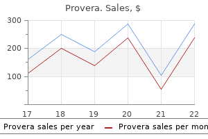
Provera
Discount 5 mg provera with mastercard
The training is implemented by PyTorch using the Adam optimizer with the learning rate initially set to be 8? The whole brain regions whose dimensions are padded to the nearest multiple of 16 are served as inputs to the modi? Fill in the holes within the tumor mask and assign voxels within the holes to necrosis area women's health clinic qe gateshead buy provera 5 mg on line. In: International Conference on Medical Image Computing and Computer-Assisted Intervention. In this paper, we present a novel three pathways U-Net structure to seg ment the brain tumor. Each modality is processed in a single pathway, flair path way for whole tumor segmentation and t1ce pathway for the enhance tumor and necrotic components segmentation. At the end of the model, these two pathways are fused together by the fusion pathway to get the final segmentation. We adopt a weighting scheme at the loss layer to alleviate the problem of label imbalance. How ever, there are multiple challenges in automatic segmentation: 1) the tumor has a low contrast with the surrounding tissue 2) large variation in tumor shape across different subjects. In the past few years, many attempts have been made to address these challenges in tumor segmentation. The convolutional neural network has become the most promising technique for image segmentation. A novel bridge strategy is used to connect the shadow layer features with the deep layer features. This bridge can help the neural network to better learn the mapping from raw data to the segmentation label and also can help to alleviate the gradient van ish problem in training stage. Necrotic components can be easily segmented from T1 post contrast image, but the peritumoral edema of the tumor is hard to be distin guished from the normal tissue. Flair image clearly shows the whole tumor but cannot distinguish necrotic components. While the T1ce image can distin guish necrotic components and enhance tumor but the peritumoral edema is hard to be distin guished from the normal tissue. Flair-pathway receives Flair image for whole tumor segmentation and t1ce-pathway receive the T1ce image for the enhance tumor and necrotic components segmentation. We fuse these two pathways together in the fusion pathway at the end of the model to get the final seg mentation. The main novelty of our method is two folds: 1) Two pathways to process different modality data. In the training stage, a 2d slices select strategy is used to prepare the training slices, and then the weights for each label is calculated by the label amount of all training slices. After the testing stage, a post-processing procedure is applied to further refine the segmentation result. T1ce-path way is used to segment the enhance tumor and necrotic components from the T1ce image, while flair-pathway is used to segment the whole tumor and brain from flair image. Both pathways use U-Net structure that can combine the low-level feature with the high-level feature by concatenating operation. For flair-pathway and t1ce-pathway, we add two auxiliary supervisions to force the network to encode more semantic con cepts, separately. Specifically, we add t1ce loss at the end of t1ce-pathway and flair loss at the end of the flair-pathway. The ground truth of the flair-pathway contains two labels, which includes the healthy brain tissue and the whole tumor. The ground truth of the t1ce-pathway contains two labels, which includes the enhance tumor and necrotic components. For fusion-pathway, the input is the sum of the last layers of flair-pathway and t1ce-pathway. The ground truth of the fusion-pathway contains all labels need to be segmented, which includes whole brain, peritumoral edema of the tumor, enhance tumor and necrotic components. For convolution operation, the kernel size is 3x3 and the step size is 1, while for deconvo lution operation, the kernel size is 4x4 and the step size is 2. The three pathways are highlighted in light-pink, light-yellow, and light-blue bands. In here, slices are selected ac cording to the number of the label in the label volume. For each slice, the number of the label (peritumoral edema of the tumor, enhance tumor and necrotic components) is counted and only the counted value large than 300 is selected. We should note that all the images are normalized to a range from 0 to 255 before training slices preparation. These three pathways segment different components of the tumor and each pathway has its unique supervision label. For flair-pathway, we segment health brain tissue and whole brain, so we set label value 4 and 1 to value 2. For flair-pathway, = {0, 2, 4}, for t1ce pathway, = {0, 1, 3}, for fusion pathway, = {0, 1, 2, 3, 4}. In the similar way, we can calculate the weights for t1ce-pathway and fusion-pathway. Specifically, only the largest vol ume is kept from the final segmentation, all other small fragments are removed. For the training dataset, we randomly select 30 subjects as the testing data, 20 subjects as the validation data and the rest 160 subjects as the training data. For the Hausdorff Distance, the value of the whole tumor is smaller on the validation dataset, but the value of enhancing tumor and tumor core is larger on the validation dataset. As we do not have the ground truth of validation data, we just show the segmentation on training dataset in Fig. Since ground truth is manually labeled, the discontinuity error might occur between adjacent slices. However, our segmentation is smoother than the ground truth, this is more biological feasible. The validation result on 30 cases of training dataset and 66 cases on the validation da taset. The green color in the whole tumor is peritumoral edema, the yellow color is the enhance tumor and the red color is the necrotic components. We can clearly see that the post-processing procedure can remove the significant mis segmentation. The comparison between before post-processing and after post-processing with ground truth. A novel weights calculation method is used to help to train the model and a post-processing procedure helps to refine the segmentation results. In the future work, we will put more efforts on improving the seg mentation ability in small regions so that the segmentation accuracy will improve in the validation dataset. Hanbury, Metrics for evaluating 3D medical image segmentation: analysis, selection, and tool. However, due to the highly heterogeneous appearance and shape, segmen tation of brain tumors is very challenging. Recent development using deep learn ing models has proved its effectiveness in the past few brain segmentation chal lenges as well as other semantic and medical image segmentation problems. Most models in brain tumor segmentation use a 2D/3D patch to predict the class label for the center voxel. U-Net is a widely used network structure for end-to-end segmenta tion and can be used on entire image or extracted patches to provide classification labels over the entire input voxels. Instead of picking the best network structure, an ensemble of multiple models, trained on different dataset or different hyper parameters, can generally improve the segmentation performance. In this study we propose to use an ensemble of 3D U-Nets with different hyper-parameters for brain tumor segmentation. In addition, we developed a linear model for survival prediction using extracted imaging and non-imaging features, which, despite the simplicity, can effectively reduce overfitting and regression errors. Keywords: Brain Tumor Segmentation, Ensemble, 3D U-Net, Deep Learning, Survival Prediction 1 Introduction Gliomas are the most common primary brain malignancies, with different degrees of aggressiveness, variable prognosis and various heterogeneous histological sub-regions, i. Due to the highly heterogeneous ap pearance and shape of brain tumors, small patches are usually extracted to predict the class for the center voxel. To improve model performance, multi-scale patches with different receptive field sizes are often used in the model [5].
Discount provera line
When polytomograms menopause fever purchase provera 5 mg on line, contrast radiography, or other special tests have been performed, copies of the reports of these tests should be obtained in addition to appropriate medically acceptable imaging reports of the skull and temporal bone. In evaluating the loss of speech, the ability to produce speech by any means includes the use of mechanical or electronic devices that improve voice or articulation. Impairments of speech may also be evaluated under the body system for the underlying disorder, such as neurological disorders, 11. How do we evaluate impairments that do not meet one of the special senses and speech listings? These listings are only examples of common special senses and speech disorders that we consider severe enough to prevent an individual from doing any gainful activity. If you have a medically determinable impairment(s) that does not meet a listing, we will determine whether the impairment(s) medically equals a listing. A mean deviation of ?22 or worse, determined by automated static threshold perimetry as described in 2. A visual field efficiency of 20 percent or less as determined by kinetic perimetry (see 2. Visual efficiency of the better eye of 20 percent or less after best correction (see 2. Disturbed function of vestibular labyrinth demonstrated by caloric or other vestibular tests; and B. An average air conduction hearing threshold of 90 decibels or greater in the better ear and an average bone conduction hearing threshold of 60 decibels or greater in the better ear (see 2. A word recognition score of 40 percent or less in the better ear determined using a standardized list of phonetically balanced monosyllabic words (see 2. The diagram of the left eye illustrates a visual field contracted to 30 degrees in two meridians and to 20 degrees in the remaining six meridians. The listings in this section describe Childhood Listings (Part B) Category of impairments resulting from respiratory disorders based on Impairments, symptoms, physical signs, laboratory test abnormalities, and response to a regimen of treatment prescribed by a treating Respiratory General Information source. Respiratory disorders along with any associated System impairment(s) must be established by medical evidence. Evidentiary Requirements pulmonary insufficiency Many individuals, especially those who have listing-level impairments, will have received the benefit of medically 3. Whenever there is evidence of such Asthma treatment, the longitudinal clinical record must include a Listing of Impairments description of the treatment prescribed by the treating source (overview) and response in addition to information about the nature and 3. The longitudinal Bronchiectasis record should provide information regarding functional recovery, if any. An individual other chronic who does not receive treatment may or may not be able to persistent show the existence of an impairment that meets the criteria of infections of the these listings. Also, the asthma listing specifically breathing includes a requirement for continuing signs and symptoms disorders despite a regimen of prescribed treatment. The most common symptoms attributable to these disorders are dyspnea on exertion, cough, wheezing, sputum production, hemoptysis, and chest pain. Because these symptoms are common to many other diseases, a thorough medical history, physical examination, and chest x-ray or other appropriate imaging technique are required to establish chronic pulmonary disease. Pulmonary function testing is required to assess the severity of the respiratory impairment once a disease process is established by appropriate clinical and laboratory findings. Gas exchange abnormalities without significant airway obstruction can be produced by interstitial disorders. Persistent hypoxemia produced by any chronic pulmonary disorder also can result in chronic pulmonary hypertension and right heart failure. Chronic infection, caused most frequently by mycobacterial or mycotic organisms, can produce extensive and progressive lung destruction resulting in marked loss of pulmonary function. Some disorders, such as bronchiectasis, cystic fibrosis, and asthma, can be associated with intermittent exacerbations of such frequency and intensity that they produce a disabling impairment, even when pulmonary function during periods of relative clinical stability is relatively well-maintained. Respiratory impairments usually can be evaluated under these listings on the basis of a complete medical history, physical examination, a chest x-ray or other appropriate imaging techniques, and spirometric pulmonary function tests. In some situations, most typically with a diagnosis of diffuse interstitial fibrosis or clinical findings suggesting cor pulmonale, such as cyanosis or secondary polycythemia, an impairment may be underestimated on the basis of spirometry alone. More sophisticated pulmonary function testing may then be necessary to determine if gas exchange abnormalities contribute to the severity of a respiratory impairment. Additional testing might include measurement of diffusing capacity of the lungs for carbon monoxide or resting arterial blood gases. In disorders of the pulmonary circulation, right heart catheterization with angiography and/or direct measurement of pulmonary artery pressure may have been done to establish a diagnosis and evaluate severity. These listings are examples of common respiratory disorders that are severe enough to prevent a person from engaging in a gainful activity. Evaluation of the impairment(s) of these individuals will proceed through the final steps of the sequential evaluation process. These disorders are evaluated on the basis of the resulting limitations in pulmonary function. Evidence of chronic infections, such as active mycobacterial diseases or mycoses with positive cultures, drug resistance, enlarging parenchymal lesions, or cavitation, is not, by itself, a basis for determining that an individual has a disabling impairment expected to last 12 months. In those unusual cases of pulmonary infection that persist for a period approaching 12 consecutive months, the clinical findings, complications, therapeutic considerations, and prognosis must be carefully assessed to determine whether, despite relatively well-maintained pulmonary function, the individual nevertheless has an impairment that is expected to last for at least 12 consecutive months and prevent gainful activity. When a respiratory impairment is episodic in nature, as can occur with exacerbations of asthma, cystic fibrosis, bronchiectasis, or chronic asthmatic bronchitis, the frequency and intensity of episodes that occur despite prescribed treatment are often the major criteria for determining the level of impairment. Attacks of asthma, episodes of bronchitis or pneumonia or hemoptysis (more than blood-streaked sputum), or respiratory failure as referred to in paragraph B of 3. Hospital admissions are defined as inpatient hospitalizations for longer than 24 hours. The medical evidence must also include information documenting adherence to a prescribed regimen of treatment as well as a description of physical signs. For asthma, the medical evidence should include spirometric results obtained between attacks that document the presence of baseline airflow obstruction. Cystic fibrosis is a disorder that affects either the respiratory or digestive body systems or both and is responsible for a wide and variable spectrum of clinical manifestations and complications. Confirmation of the diagnosis is based upon an elevated sweat sodium concentration or chloride concentration accompanied by one or more of the following: the presence of chronic obstructive pulmonary disease, insufficiency of exocrine pancreatic function, meconium ileus, or a positive family history. The quantitative pilocarpine iontophoresis procedure for collection of sweat content must be utilized. Two methods are acceptable: the "Procedure for the Quantitative Iontophoretic Sweat Test for Cystic Fibrosis" published by the Cystic Fibrosis Foundation and contained in, "A Test for Concentration of Electrolytes in Sweat in Cystic Fibrosis of the Pancreas Utilizing Pilocarpine lontophoresis," Gibson, I. The nonpulmonary aspects of cystic fibrosis should be evaluated under the digestive body system (5. Because cystic fibrosis may involve the respiratory and digestive body systems, the combined effects of the involvement of these body systems must be considered in case adjudication. The results of spirometry that are used for adjudication under paragraphs A and B of 3. Two of the satisfactory spirograms should be reproducible for both pre-bronchodilator tests and, if indicated, post-bronchodilator tests. Wheezing is common in asthma, chronic bronchitis, or chronic obstructive pulmonary disease and does not preclude testing. The effect of the administered bronchodilator in relieving bronchospasm and improving ventilatory function is assessed by spirometry. If a bronchodilator is not administered, the reason should be clearly stated in the report. Pulmonary function studies performed to assess airflow obstruction without testing after bronchodilators cannot be used to assess levels of impairment in the range that prevents any gainful work activity, unless the use of bronchodilators is contraindicated. Post-bronchodilator testing should be performed 10 minutes after bronchodilator administration. If the spirogram was generated by any means other than direct pen linkage to a mechanical displacement-type spirometer, the testing device must have had a recorded calibration performed previously on the day of the spirometric measurement. If the spirometer directly measures flow, and volume is derived by electronic integration, the linearity of the device must be documented by recording volume calibrations at three different flow rates of approximately 30 L/min 3 L/6 sec) 60 L/min 3 L/3 sec), and 180 L/min 3 L/sec). If reproductions of the original spirometric tracings are submitted, they must be legible and have a time scale of at least 20 mm/sec and a volume scale of at least 10 mm/L to permit independent measurements. A diffusing capacity of the lungs for carbon monoxide study should be purchased in cases in which there is documentation of chronic pulmonary disease, but the existing evidence, including properly performed spirometry, is not adequate to establish the level of functional impairment. At altitudes above sea level, the inspired O2 concentration may be raised to provide an inspired O2 tension of approximately 150 mm Hg. Abnormal hemoglobin or hematocrit values, and/or carboxyhemoglobin levels should be reported along with diffusing capacity. The ability of the individual to follow directions and perform the test properly should be described in the written report.
Syndromes
- People who were young (less than 30 years) when the disease started
- Numbness of hands or feet
- Pain during urination (dysuria)
- Injury to the lung, which could cause more breathing problems
- For other types of stings/bites, you may be told to apply vinegar or a meat tenderizer/water solution to neutralize the venom.
- You may need stool softeners to avoid constipation.
- MRI of the brain
- Blood tests to check for problems and degree of poisoning, including calcium level
- Is the child easily distracted?
- Getting full quickly when eating
Purchase provera with amex
Chapter 8 | Cardiac Arrest | 111 Box 8-1 | Reversible Causes of Cardiac Arrest: Hs and Ts Practice Note Hs Ultrasonography can be a useful tool for recognizing Hypovolemia several underlying causes of cardiac arrest menopause question buy provera 5mg online, including Hypoxia pulmonary embolism, tension pneumothorax, Hydrogen ion excess (acidosis) cardiac tamponade and hypovolemia. Consider use Hyper or hypokalemia of ultrasonography if the equipment and a skilled Hypothermia technician are readily available, and if doing so will not Ts impede or delay resuscitation efforts. A fuid challenge can aid in determining underlying causes of the cardiac arrest include a 12 or whether hypovolemia is contributing to the cardiac arrest. Suspect acidosis in patients with diabetes or probable acute or chronic renal failure. In patients Tamponade with metabolic acidosis, the administration of an initial dose of sodium bicarbonate (1 mEq/kg) may Cardiac tamponade occurs when fuid accumulates be indicated. Sodium bicarbonate, if used, should be in the pericardial sac, compressing the heart and administered early in conjunction with standard cardiac preventing it from pumping effectively. Pre-arrest physical examination fndings may include the Potassium imbalances can precipitate cardiac arrest. Sodium Treatment is pericardiocentesis (needle aspiration of bicarbonate is the preferred method of addressing fuid from the pericardial sac). Suspect hypokalemia in patients with dehydration Tension pneumothorax occurs when air accumulates in or overuse of diuretics. Compression of the vena cava is intravenous administration of a dilute solution of leads to impaired venous return and decreased cardiac potassium chloride. Penetrating chest trauma is a common cause of tension pneumothorax, but the condition can also Hypothermia develop in older patients with underlying lung disease and in patients who smoke. For patients with severe hypothermia (body temperature Pre-arrest physical examination fndings may include less than 86 F [30 C]) and cardiac arrest, core hypotension, tachycardia, absent breath sounds on the rewarming (with cardiopulmonary bypass, extracorporeal affected side, jugular venous distension, hyperresonance blood warming with partial bypass or thoracic lavage on percussion and tracheal deviation away from the with warmed fuids) is indicated. Diffculty ventilating the warmed humidifed oxygen may be administered as patient may also be a sign of tension pneumothorax. Initial treatment is with needle chest decompression or Overdoses (of both illicit and therapeutic drugs) thoracostomy. Drugs that are frequently implicated in cardiac arrest include cocaine, methamphetamines, opioids (heroin, fentanyl), Thrombosis (Pulmonary Embolism)? Reversal agents are specifc to In massive pulmonary embolism, obstruction of the pulmonary artery and the release of vasoconstrictive 114 | American Red Cross | Advanced Life Support mediators from the thrombus lead to cardiogenic shock, potentially helpful, are secondary interventions and have which can quickly lead to cardiac arrest. Conditions not been proven to lead to improved survival rates or in the patient history associated with prolonged neurologic function in patients who experience cardiac immobilization or venous stasis, hypercoagulability or arrest. Witnessed cardiac arrest and respiratory distress for at least 2 minutes (in the case of a nonshockable before arrest also may point to pulmonary embolism as rhythm). Thrombosis (Myocardial Infarction) Practice Note Myocardial infarction can lead to cardiac arrest. Approach to the Patient the Cardiac Arrest: Adult Treatment Guideline Practice Note summarizes the approach to a patient in cardiac arrest. Remember that when an advanced airway is in place, Assess and Recognize chest compressions are performed continuously without pausing to deliver ventilations. Ventilations After determining that the patient is not responsive, are delivered at a rate of 1 ventilation every 6 not breathing and has no pulse, call for assistance and seconds. Shockable Rhythms Care Ventricular fbrillation and pulseless ventricular tachycardia require defbrillation as soon as possible. Medications, although Chapter 8 | Cardiac Arrest | 115 to chest compressions; Figure 8-5) to determine next the energy dose depends on whether the defbrillator actions: is biphasic or monophasic. Practice Note If defbrillation is initially successful in terminating Check the pulse only if an organized rhythm is the cardiac arrest rhythm but ventricular fbrillation or present. Practice Note Always precede the delivery of a shock by announcing the intention to shock in a clear, succinct manner. Before During rhythm and pulse checks, pause delivering a shock, perform a visual scan to ensure that compressions for no more than 10 seconds. Administering shocks establishes a temporary ?blank If the rhythm check reveals a shockable rhythm, slate by eliminating all electrical activity in the heart (in resume compressions as soon as the charging other words, it temporarily induces asystole). Medications Various medications may be used in the treatment of ventricular fbrillation or pulseless ventricular tachycardia. The vasoconstrictive and positive ionotropic effects Figure 8-5 | Minimize pauses in compressions to less than 10 seconds during rhythm and pulse checks. In addition, it is extremely important to look for and address potential underlying causes of the cardiac arrest. Figure 8-6 | Ensure that all providers are clear of the patient and the bed before delivering a shock. When delivering the Terminating the shock, face the team, rather than the defbrillator. However, in some Practice Note situations it may be appropriate to consider prolonging the resuscitation effort, using specialized interventions or Choose one antiarrhythmic agent (amiodarone both. For example, it may be appropriate to prolong the or lidocaine) and use it for the duration of the resuscitation effort when more time is needed to address resuscitation effort. Do not alternate between the underlying cause of the cardiac arrest (for example, the two. Deoxygenated blood is Magnesium sulfate should not be administered too removed from the body, passed through a membrane quickly because it may induce hypotension. This is largely a result of the global consequences of hypoxemia and the ischemia/reperfusion response, in addition to the precipitating cause of the cardiac arrest itself. Blood fow must be restored to tissues that have been deprived of oxygen in order to prevent tissue death; however, reperfusion of previously ischemic tissues can induce an infammatory response that causes cellular injury in addition to that caused by the ischemia itself. Figure 9-1 | Expert care during the immediate post?cardiac Sometimes referred to as post?cardiac arrest arrest period can improve outcomes for the post?cardiac syndrome, the pathophysiologic consequences of arrest patient. Brain injury, caused by ischemia and capnography and noninvasive blood pressure monitoring cerebral edema, is a signifcant cause of morbidity or arterial pressure monitoring as needed. The ischemia/reperfusion response can trigger a systemic infammatory History response, which can lead to multiple organ dysfunction. The underlying cause of the cardiac arrest may continue Physical Examination to have pathophysiologic consequences during the Conduct a focused cardiopulmonary physical post-arrest period. The Post?Cardiac Arrest Care: Adult Treatment Guideline summarizes the approach to caring for a Optimizing Ventilation and Oxygenation patient during the immediate post?cardiac arrest period. Provide high-fow supplemental oxygen until the oxygen saturation can be measured, and then provide Assess and Recognize the minimal level of supplemental oxygen needed to maintain an oxygen saturation of at least 94%. Support ventilations, starting at a rate of 10 breaths per Establishing cardiac monitoring as needed. Hypocapnia can and arterial blood gases are used to guide ventilatory decrease cerebral blood fow and worsen neurological management. Continuously monitor the patient using Establishing noninvasive blood pressure monitoring or arterial pressure monitoring to assist in managing capnography and pulse oximetry to ensure optimal hemodynamics. Blood pressure can be extremely labile during the Care post?cardiac arrest period. In general, hypoglycemia (blood glucose anuric patients) or a pulmonary artery catheter (if one is < 80 mg/dL) should be treated with dextrose, and in place). Each year in the United States, an estimated 660,000 people require hospitalization for myocardial infarction related to coronary artery disease. Classifcation of Acute Manage complications, such as ischemia-induced Coronary Syndromes arrhythmias that can lead to cardiac arrest and death. For example, patients with Clinical Presentation diabetes may experience ischemia without pain (?silent ischemia?). Cardiac troponin T (cTnT) and cardiac troponin I (cTnI) biomarkers are preferred for diagnosing myocardial injury. For diagnosis, it is important to assess both the peak troponin level and changes in troponin level over time, so cardiac troponin levels should be measured at initial presentation and then 3 to 6 hours later. For high-sensitivity markers, some protocols suggest measurement at initial presentation and then 2 hours later. In addition to being prognostic (for both short and long-term outcomes), cardiac troponin levels in combination with risk stratifcation are useful to guide treatment decisions. Patient Assessment Chest pain or discomfort results in approximately 7 million emergency department visits each year. To expedite appropriate therapy for all patients presenting with chest pain or discomfort, providers must be able to rapidly discern the underlying cause.
Discount 10mg provera visa
The decision as to whether to give post-operative radiotherapy should be made based upon the presence or absence of adverse pathological features menstruation quality 5 mg provera for sale. However, for early vulvar disease a sub group has been identified in whom radiotherapy is recommended. However, according to some gynaecological oncologists, there is no evidence to support the pathological parameters quoted in the guidelines. These experts state that evidence for post-operative radiotherapy exists only in patients with > 1 macro-metastasis, > 2 micro metastases, extranodal spread or margins < 5mm. These patients are considered to be at intermediate or high risk for recurrence and significant failure rates occur in the absence of radiotherapy (75) (85) (86) (87). They published incidence proportions for ?low, ?intermediate and ?high risk as 67%, 18% and 15% respectively. Intermediate and high risk patients are defined as patients with 2 unilateral lymph nodes, tumours of > 2 cm with 1 positive node and lesions > 8 cm with no positive nodes. Incidence of post-surgical recurrence the treatment of recurrent vulvar cancer has not been standardized due to the various ways in which recurrence may present. Two expert reviewers in this report both suggested that surgery is almost always the treatment of choice for local recurrence but nodal recurrences will almost always require radiation (either alone or in combination with surgery). Three patients developed local recurrence and all were successfully salvaged with further radical surgery. The development of distant metastases is rare without local recurrence and therefore a branch on the decision tree has been omitted. The omission of a branch on the decision tree corresponding to those who did not receive radiotherapy at initial treatment or at recurrence, and who subsequently developed isolated distant recurrence amenable to palliation with radiotherapy (such as brain or bone metastases) is unlikely to alter the overall estimate for optimal radiotherapy utilisation. Sensitivity Analysis Sensitivity analysis allows the assessment of the impact of changing the value of the variables on the overall end result. For the gynaecological decision tree, one data item was identified as being uncertain. In endometrial cancer, no data could be identified to estimate the proportion of early stage endometrial cancer patients who undergo a lymph node dissection. To assess the impact of this uncertainty on the overall estimate of the need for radiotherapy in all gynaecological cancers, a sensitivity analysis was performed for each of the variables. Once the decision trees for all tumours are completed, a tornado analysis will be performed whereby the impact that each of these variables has on the overall estimate of the proportion of cancer patients needing radiotherapy will be examined. The graphs below show that the optimal proportion of gynaecological cancer patients who should receive radiotherapy based on evidence and incidence of attributes for radiotherapy is 35%. This proportion could vary from a low of 31% to a high of 39% depending on the proportion of patients undergoing endometrial cancer surgery who also have a lymph node dissection (varied between the extremes of 10% to 90%). Tornadodiagram of sensitivityanalysisforgynaecologicalcancer T ornado Diagram at G ynaecancer proportionofendometrialcancerpatientsundergoingnodedissection:0. The optimal utilisation rates for the five gynaecological tumour sub-sites are shown in Table 8. Table 8: Optimal radiotherapy utilisation rates by gynaecological sub type Tumour Sub % of Overall optimal Proportion of all site gynaecological radiotherapy utilisation rate cancer patients cancer for sub-site (%) that should receive radiotherapy Cervix 23 58 0. Consensus Statement: National Institutes of Health consensus development conference statement on cervical cancer. The palliation of brain metastases in a favorable patient population: a randomized clinical trial by the radiation therapy oncology group. Multivariate analysis of the histopathologic prognostic factors of cervical cancer in patients undergoing radical hysterectomy. Early invasive carcinoma of the cervix (3 to 5 mm invasion): risk factors and prognosis. Definition and prognostic significance of microinvasion in the uterine cervix squamous lesion. A reappraisal of the International Federation of Gynecology and Obstetrics staging system for cervical cancer. Prognostic value of performance status assessed by patients themselves, nurses, and oncologists in advanced non-small cell lung cancer. Concurrent radiation and chemotherapy for carcinoma of the cervix recurrent after radical surgery. The American Brachytherapy Society recommendations for high-dose-rate brachytherapy for carcinoma of the endometrium. Risk factors for recurrence in clinically early endometrial carcinoma: an analysis of 183 consecutive cases. Recurrent adenocarcinoma of the endometrium: a clinical and histopathological study of 379 patients. Significance of true surgical pathologic staging: a Gynecologic Oncology Group study. The role of adjuvant radiotherapy in carcinoma of the endometrium results in 550 patients with pathologic Stage I disease. Excellent long-term survival and absence of vaginal recurrences in 332 patients with low risk stage I endometrial adenocarcinoma treated with hysterectomy and vaginal brachytherapy without formal staging lymph node sampling: report of a prospective trial. The relationship of local and distant failure from endometrial cancer: defining a clinical paradigm. Recurrent endometrial cancer after surgery alone: results of salvage radiotherapy. Postoperative external irradiation and prognostic parameters in stage I endometrial carcinoma: clinical and histopathologic study of 540 patients. Surgery and postoperative radiotherapy versus surgery alone for patients with stage-1 endometrial carcinoma: multicentre randomised trial. Postoperative radiotherapy and surgery in Stage I endometrial carcinoma: a 10-year experience. Radiation therapy versus pelvic node resection for carcinoma of the vulva with positive groin nodes. Groin dissection versus groin radiation in carcinoma of the vulva: a gynecologic oncology group study. Radiation therapy in management of carcinoma of the vulva with emphasis on conservation therapy. Radical resection of vulvar malignancies: a paradigm shoft in surgical approaches. Management of regional lymph nodes and their prognostic significance in vulvar cancer. Genitourinary Cancers (excluding prostate cancer) Renal Cancer Table 1:R enalC ancer. Th e incidence ofattributes used to define indications forradioth erapy K ey Populationor Attribute Proportionof Q ualityof R eferences N otes subpopulationof populationwith inform ation interest thisattribute A Allregistrycancers R enalcancer 0. Indications for radiotherapy the primary treatment modality for renal cancer is radical surgical resection. The decision tree has not included positive margins as a reason for giving radiotherapy. There are also other less common clinical scenarios that may be considered for radiotherapy such as symptomatic lung metastases. However, these situations are rare enough that their omission from the tree is unlikely to have a significant impact on the overall radiotherapy utilisation rate for renal cancer. Overall incidence Renal cancer comprises 3% of all cancers according to the Australian Institute of Health and Welfare statistics for 1998. Stage proportions the treatment of renal cancer is predominantly radical surgery (total or partial nephrectomy) in patients with no metastatic disease. Surgery is also sometimes indicated in patients with limited metastatic disease (16), (15). The proportion of patients diagnosed with M1 disease at initial presentation, according to the South Australian Hospital Registry, is 31% (5). By excluding the data on unstaged patients, patients with metastatic disease represent 27%, which is similar to the South Australian figure. Operability rate Not all patients with M0 disease will be fit enough for a radical nephrectomy. No direct accurate data on performance status or incidence of co-morbidities in renal cancer were available.

Buy 10mg provera visa
Until having high level classifcation accuracy women's health clinic reading pa discount provera 10mg online, convolution and pooling layers are arranged. In addition, some feature maps can be found in each convolutional layer and weights of convolutional nodes in the same map are shared. These arrangements allow for the learning of diferent characteristics of the network while keeping the number of traceable parameters. C1 S1 C2 S2 feature image feature maps feature maps feature maps 28x28 14x14 10x10 14x14 Input image 5x5 2x2 5x5 2x2 32x32 convolution subsampling convolution subsampling Feature Extraction Classification Figure 1. First Structure: No learning activity takes place in this part; in other words, no weight updating is calculated. Second Structure: the extracted features from the input images, which belong to training set, are acquired via combining these features on a matrix. In this study, in order to reduce the noise, nonlocal means and local smoothing methods were used [19]. Some important structures and details in an image can behave like noise; these important details may also be removed. A sample original image is shown in Figure 4a and a preprocessed image is shown in Figure 4b. Watershed built on the terms of watershed lines is connected to topography and water source basin. At the beginning of recursionXh basin cluster was counted as equal to dot clusters that have hmin value. This info is calculated by diferentiating the frst derivate of the change between pixel values. For segmentation of an image, diferent pointer pixels belonging to each aimed class are needed. Watershed transformation has interesting features that are useful during image segmentation applications in mathematical morphology. In this study, in order to avoid excessive segmentation, pointers were gathered initially by the assistance of morphological operators and then the watershed algorithm was applied to the image that includes pointers. Opening Process: Objects in the image and gaps among them are cleaned according to the size of the structural element. Objects that stay on the upper side of the image shrink compared to the size of their original image. Closing Process: Dots in the image are close to each other at the end of closing process. As in the expansion process, dots that were close to each other united and gaps between them were flled. Afterwards, the soft tissue of the brain was obtained by extracting the skull image from the real image, as shown in Figure 4d. Detection of the tumor was conducted by morphological procedures and suitable threshold values, as shown in Figure 4e. Used benign and malignant tumor images belonging to each axial, coronal, and sagittal plane were digitized at 256? Nine patients images were used for training and 7 patients images were used for testing. In the preprocessing stage, denoising and normalization operations were employed in order to prepare the input images for the next stage. The weights between the hidden layer and the output layer were calculated analytically by using the least square method. Based on the extensive experiments, the convolution flter size was determined as 5, the K value was chosen as 16, and the pooling size was chosen as 3. In addition, for identifying the most appropriate C value, an interval search was performed among 2? Convolution and pooling process results are shown in Figure 6 and Figure 7, respectively. The second layer is the convolution layer following the input layer, where six convolution flters are used. The third layer is the pooling layer that was employed after the frst convolution layer. The fourth layer is another convolution layer where 12 convolution flters are used. Gabor wavelet transform and statistical based studies, all features were extracted from the tumor or tumor like portion. Statistical features-based method and Gabor-wavelet features-based method yielded accuracy values of 96. Thus, it can be concluded that deep learning-based methods outperform the methods compared. Malignant tumors are denoted as positive and benign tumors are denoted as negative. There were 5 max pooling layers in the architecture, which follow some of the convolution layers. The second convolution layer flters the output of the previous layer by using 256 flters. The fourth and ffth convolutional layers have the same structure of the third convolutional layer. As we mentioned earlier, three fully connected layers follow the convolutional layers. There are two dropout layers, which come after frst and second fully connected layers, with probability of 0. The raw input directly applies to the network for learning of spatial correlations in natural images. Based on the extensive experiments, the convolution flter size was set to 5, the K value was chosen 16, and the pooling size was chosen 3. Deep feature learning with discrimination mechanism for brain tumor segmentation and diagnosis. The application of convolution neural networks in handwritten numeral recognition. Journal of the Faculty of Engineering and Architecture of Gazi University; 2016; 31: 737-747 (in Turkish). Mathematical morphology and geology: when image analysis uses the vocabulary of earth science a review of some applications. In: International Conference on Computer Analysis of Images and Patterns; 22-24 August 2017; Ystad, Sweden. Effects include increased cancer risk, cellular stress, increase in harmful free radicals, genetic damages, structural and functional changes of the reproductive system, learning and memory deficits, neurological disorders, and negative impacts on general well-being in humans. Damage goes well beyond the human race, as there is growing evidence of harmful effects to both plant and animal life. Cell phone use and risk of thyroid cancer: a population-based case-control study in Connecticut. Abstract-Brain tumor is a group of tissue that is prearranged brain tumor using image processing techniques. It occurs when cell get detection of a brain tumor at an early stage is a key issue for abnormal formation within the brain. The seriousness of brain clinically suspected, radiological evaluation is required to tumor is very big among all the variety of cancers, so to save a life immediate detection and proper treatment to be done. It is very essential to compare brain therapy, surgery, radiation, or chemotherapy, is decided. It is very difficult to have vision is evident that the chances of survival of a tumor-infected about the abnormal structures of human brain using simple patient can be increased significantly if the tumor is detected imaging techniques. As a result, the study of most influential development in Data Mining and Machine Learning in the past decade. They combine multiple models into brain tumors using imaging modalities has gained one usually more accurate than the best of its importance in the radiology department. Ensemble methods combine the procedure of neural brain tumor identification is done by an image processing. Preprocessing, segmentation, feature extraction, and image data from the collection of database using median classification. Secondly segmentation is performed by using filtering, second stage is segmentation using Fuzzy C-means clustering algorithm.
Order 5 mg provera with amex
Such testing is straightforward for simple become familiar enough with the type of applicator and the devices such as manual afterloading sources menopause and sexual dysfunction provera 10mg lowest price, but can be very possible errors involved with small deviations from the ideal complex and model speci? After the pro tance testing is an opportunity for the physicist to learn in cedure, if there is the possibility of more than a 10% error in detail the operational characteristics of the system and cor Medical Physics, Vol. Procedure End point Frequency Procedure End point Frequency evaluate dimensions/ source identity initially evaluate spacing and ribbon geometry and seed initially serial number physical length and diameter no. During acceptance A number of useful references are available for designing testing all clinical procedures associated with device should a program to con? This is also a good time to develop training and 40 outline a number of basic tests and give recommen programs for dosimetrists, technologists, and others who will 31,97 dations as to frequency. Annual calibra nonvolatile form which is heavily encapsulated in stainless tion checks of all long-lived sources are recommended steel. Remote afterloading brachytherapy devices thin, easy-to-rupture titanium tubing for interstitial brachy As with any treatment delivery system, functional remote therapy. Such sources must be handled very carefully to pre 125 125 afterloading quality assurance tests should anticipate the vent leakage of I. There are three principal to be leak tested prior to use in a second patient or after form quality assurance end points: accuracy of the source selec intensive handling or manipulation. In Dosimetric evaluation of intracavitary applicators is not addition, all remote afterloaders have error and malfunction straightforward as accurate measurement of brachytherapy detection systems ~?interlocks?! However, when adopt signed to retract all sources and sound an alarm when the ing sources and applicators that differ signi? Alternatively, Monte Carlo photon transport calcu order in which equal strength radioactive spheres of lations can be used if the source/applicator geometry is 137Cs or 60Co and geometrically identical spherical spac known. Different treatment times may be programmed program should have three testing frequencies: initial accep for each channel. Fixed source-train devices have no capability of compos depth of a dwell position sequence is continuously ~or ing arbitrary source trains from elemental components nearly so! Single stepping source devices consist of a single cable driven high intensity source, which moves from each sources. Additional core quarterly quality assurance tests for a remote afterloading facility. The dose per pulse is chosen tests at three frequencies: daily, quarterly, and annually/ so as to duplicate, on average, the hourly dose rate charac initially. Additional commissioning and annual quality assurance tests for a remote afterloading facility. Such tests should be completed before be check of source radiation output and timer accuracy for ginning applicator insertion in the? These tests greatly 60-s programmed dwell time to the expected value, an over Medical Physics, Vol. A method of reconstructing the three-dimensional geom 97 ~see Williamson for speci? At the very least, the inherent posi more advanced features such as catheter-trajectory re tional accuracy of the machine should be tested since the construction algorithms and a menu of algorithms for source has been changed. A means of assigning the source type, strength, and treatment time ~or dwell time! Su be comprehensive, approaching the thoroughness of initial perposition is then used to calculate the multiple source acceptance testing in this regard. Conventional systems require the user more comprehensively ~although measurement over range of to heuristically evaluate the dose distribution by exam use may be practical!. Positional accuracy should be checked ining isodose curve distributions in manually selected carefully, including the condition and dimensions of all planes. The radioactive source locations source remote afterloaders have more advanced features should be compared directly to their dummy source counter such as dwell-weight optimization algorithms, dose vol Medical Physics, Vol. Prevention of software related treat ment errors, as well as data entry errors of human origin. Medical health physics Dose calculation algorithms should be tested both to verify that the algorithm executes as speci? These include manual loading of sources for intrac or volumes, prescribing dose to the tumor and normal tis avitary therapy, manual loading for interstitial therapy, re sues, computerized treatment planning, and treatment deliv mote afterloading for low-dose rate intracavitary or ery. The individual functions of brachytherapy team mem interstitial therapy, stereotactically guided procedures for bers are rooted in the training and education of the particular treating brain lesions, ultrasonically guided procedures for team member. However, the effective and safe use of treating prostatic cancer, and eye plaques for treating ocular brachytherapy depends on a thorough understanding of the tumors. Since each requires specialized equipment, a com science of all aspects of isotope treatment including the roles plete brachytherapy program is very expensive, and gener of all team members. The physician Most institutions limit their brachytherapy programs to one needs to be aware of dose distribution around different or a few of the above techniques. This section lists the equip source arrangements to adequately prepare for an implant. The list the physicist needs to be knowledgeable of anatomy such starts with a set of equipment for radiation safety applicable that the relationships between tumor volume and surrounding to all techniques, and necessary even if only one technique is normal tissues are considered during the treatment planning used. This give-and-take is crucial to the planning, execu tion, and delivery of effective and safe brachytherapy. Equipment needed for any and all brachytherapy proce physics is not on-site for pre-treatment planning, simulation dures: Medical Physics, Vol. Nuclear Regulatory Commission, Title 10, Chapter 1, Code of Federal Regulations-Energy, Part 20, ?Standards C. Nuclear Regulatory Commission, Guide for the wire cutters, Preparation of Applications for Medical Use Programs, miscellaneous buttons, portable soldering iron, etc. Nuclear Regulatory Commission, Quality Manage ment Program and Misadministrations, Federal Register D. Nuclear Regulatory Commission, Quality Manage ultrasound system with brachytherapy software; ment Program, Regulatory Guide 8. Nuclear Regulatory Commission, Information Re personal computer with eye plaque software or a calcula quired for Licensing Remote Afterloading Devices, Policy tor. Selbach, ?Reference air kerma can Association of Physicists in Medicine ~American Institute of Physics, rate determination of an iridium-192 brachytherapy source, in Quality New York, 1993!. Khan, ?Monte Carlo evaluation 46, American Association of Physicists in Medicine ~American Institute of the Sievert integral for brachytherapy dosimetry, Phys. Klevenhagen, ?An experimental and calculated surance ~American College of Radiology, Philadelphia, 1990!. Williamson, ?Monte Carlo and analytic calculation of absorbed dose 6American College of Medical Physics report, Radiation Control and 137 near Cs intracavitary sources, Int. Williamson, ?The Sievert integral revisited: evaluation and exten ?Consensus guidelines for high dose rate remote brachytherapy in cervi 125 169 192 sion to I, Yb, and Ir brachytherapy sources, Int. Williamson, ?Practical quality assurance for low dose-rate brachy standards, Brachytherapy Physics, edited by J. Meigooni, ?Dosimetry of interstitial brachytherapy sources: Rec tional Brachytherapy Programme and Abstracts. Williamson, ?Experimental validation of Monte Carlo dose nium capsule of the clinical I-125 seed, Med. Dennett, ?Dose calculation and measurements for kerma rate with large volume spherical ionization chambers, Phys. Dale, ?A Monte Carlo derivation of parameters for use in the tissue for 125-I stereotactic brain implants, Int. Bassano, ?Lung density effect on 125I distribu erized optimization of 1251 implants in brain tumors, Int. Ragde, ?Brachytherapy and organ brachytherapy sources in a heterogeneous phantom with cylindrical sym metry, Med. Anderson, Atlas of Brachytherapy ~Mac sources in the presence of bounded heterogeneities, Int. Presser, ?Classical systems for temporary in terstitial implants, in Modern Clinical Brachytherapy Physics, edited by ity, in Modern Clinical Brachytherapy Physics, edited by J. Cevc, ?Dimension averaging, a simple method for for idealized implant geometries, Med.
Rorippa armoracia (Horseradish). Provera.
- Are there safety concerns?
- Urinary tract problems, fluid retention (edema), cough, bronchitis, achy joints and muscles, gout, gallbladder disorders, sciatic nerve pain, colic, intestinal worms in children, and other conditions.
- How does Horseradish work?
- Are there any interactions with medications?
- Dosing considerations for Horseradish.
- What is Horseradish?
Source: http://www.rxlist.com/script/main/art.asp?articlekey=96281
Purchase provera toronto
For a review of external beam photon dose calculations women's health clinic gadsden al provera 10mg discount, characterization from source characterization see Ahnesjo and Aspradakis (1999). As long as a successful and reproduc information ible system is unchanged, it can be safely handled by empirical knowledge. The output in terms of calculated doses relies etry is of utmost importance when clinical knowledge obtained on the calculation algorithms, and the type of input includes the radia with one system is to be transferred into another. If the electron ranges Coherent/total are short compared to the dimensions of voxels used in treat 0. The relationship between absorbed dose in water according to the contributions from photoelectric absorption, in a point P from primary photons, Dprim, and collision kerma, coherent, and incoherent scattering as a function of photon energy E. The absorbed dose absorption and linear attenuation coefcients, respectively, is a contributed by subsequent generations of photons, that is, ?the measure of the average amount of energy that is transferred to scatter dose, depends on the full 3D environment surrounding the secondary photon in a single photon interaction (Table 11. This is caused been developed and applied for calculations around 192Ir sources by the scattering process being relatively elastic. It was later extended to perform 2D fractional energy loss) in this energy interval. On the An analytical method using precalculated scatter-to-primary other hand, the secondary electron range is considerably shorter ratios was used to account for limited phantoms and heteroge than the photon mean free path and can be considered to deposit neities around 192Ir sources (Anagnostopoulos 2003). Poon and Verhaegen developed correction-based methods intended for use with 192Ir sources to take into account lack of scatter in breast implants (Poon and 11. The frst-principle methods represent general approaches that perform transport calculations in the actual media and account 11. The description below concentrates total doses, due to the presence of high atomic number shields on applications in treatment planning. The approach is correct for the dose contributed Methods and cc Method by primary photons but not for scattered photons. It can be a Superposition/convolution methods are based on superposing reasonable approximation at short distances from 137Cs and192Ir the distribution of energy released by a group of photons with sources, where doses from primary photons dominate. A scatter separation method that accounts for suitable lattices of transport lines (computational grid) over the the location of heterogeneities but not for their lateral extent was calculation volume. The scatter-to-primary in two subsequent steps using a ?successive scattering approach ratio in low-Z materials at 60 keV is around 80%. The individual (Carlsson and Ahnesjo 2000a) designed to minimize system lead shields and bulky high-Z 241Am sources presented a chal atic errors due to kernel discretization and approximations in lenging and truly 3D problem that would clearly beneft from multiple-scatter dose. Phase space fles are large and thus difcult to dis application in treatment planning requires fast calculations and tribute and commission. A rationale Z3, low-energy photon transport calculations are very sensitive Model-Based Algorithms Doing Nonwater Heterogeneity Corrections 151 to material composition. Tese external beam modalities also beneft from more derived by earlier simpler algorithms for which clinical experi detailed tissue composition information due to the strong rela ence existed. Tus, diferences between account only for the efect of fnite patient dimensions and appli values of dose to medium and dose to water reported in these cator components/shields. Concerns treatment planning being performed in a grid with voxel dimensions of 3 the dose to bulk relating to the conversion at least 1 mm and the average bulk medium composition. Hypothesizing the cell absorption coefcients to convert dose-to-medium into dose nuclei (n) to be the radiation target, the aim was to study which to-water. More knowledge on the diferences between dose-to of Dw,m and Dm,m come closest to Dn,m. Under the investigated medium in medium and dose-to-water in medium under various assumptions, Dw,m was found to be a good substitute for Dn,m for assumptions of the size of the water cavity would be valuable. Further studies on the topic are warranted and can be expected in the close future. The notation for absorbed dose estima corrections on calculated tion at location r is Dx,y? Z(r), where x denotes the choice of dose Doses?clinical implications specifcation medium (either water or local medium at r); y denotes the voxel composition used for the radiation transport In a Vision 20/20 article, Rivard et al. This manuscript presented the results in terms of high for applicators and air?tissue interfaces, or applicators and air?. In function of photon energy for various materials found in applicators addition to the review presented in the following, the interested and source compositions. More recently, clinical confgurations have been short, the radial dose function decreases quickly, and the con studied using increasing medium complexity. In the following tribution of primary photons becomes less important compared sections, a treatment site-specifc review is given. Taking into account the impor in radiation therapy, the size of the sources, typically 0. Still Meigooni and sources are not very forgiving of any type of heterogeneities, as Nath (1992) reported a possible dosimetric impact of up to 6% illustrated in Figure 11. Teir results were lead to D90 variations of 2% to 3% depending on the seed model expected based on fundamental physics cross-section behavior. A new, However, these were for single source confgurations in geom water-equivalent seed model was proposed in the same work etries where uniform water was replaced by a uniform medium (Afsharpour et al. Of course, these are average variations 154 Comprehensive Brachytherapy over the whole organ. Local dose variations in close proximity and concluded that the expected variation within the popula to seeds are much higher at tens of percent (Chibani et al. Retrospective dose calculations of a The reader should note that contrary to permanent prostate clinical cohort by Carrier et al. The efect of calcifcation within the prostate gland was carrier (Silastic) of eye plaque applicators have a large efect on investigated by Chibani and Williamson (2005). An improved characterization applicators by 8% to 10% at 1 cm and up to 15% at 2 cm or of of the efect of localized calcifcation from clinical image sets axis (Chiu-Tsao et al. Dose to critical ocular structures was approximately 21 keV, although eBx is an x-ray source and has a also shown to be 16% to 50% less than expected compared with broad, fltered spectrum (Liu et al. Tese values are radio agent, which leads to an attenuation of about 6% (Mille et al. This is because the breast composition, a mix of-axis dose can be reduced by as much as 90% (Tomson et al. As such, dose diferences magnitude to that for permanent prostate implants at about 2% around a single seed embedded in breast tissue can lead to dose (Tomson et al. For clinical breast multiseed implant confgurations, 92% dose reduction for the lacrimal gland. Furthermore, Rivard taking into account the tissue composition impact on clini et al. Note that for most of the abovementioned adipose tissue would lead to increased dose to the skin by as works, the eye was considered to consist of water, and the hetero much as 25% due to the lower attenuation of the adipose tissue. Only the work by Tomson and Rogers (2010) looked Model-Based Algorithms Doing Nonwater Heterogeneity Corrections 155 at the efect of using realistic eye tissue compositions. At lower energy, namely, that of 3% relative to water, and the eye lens dose is about 7% to 9% 169Yb, Lymperopoulou et al. Some leave air in the balloon, while oth received sublobar resection in stage I nonsmall cell lung cancer. The seeds are weaved into as a contrast agent for the MammoSite balloon (Hologic Inc. The diference relative to a water-only geometry set to cover the staple line plus a 2-cm margin and to deliver a showed variations depending on the balloon diameter (4, 5, and prescription dose of 100 to 120 Gy at 0. The seeds are placed 1 cm apart in a strand vious studies using a diferent geometry. Two of such strands 169 showed that the contrast has a larger efect on lower energy Yb are used and afxed 0. This was further illus comparing various confgurations from single to multidwell trated in a retrospective dose calculation study of a few clinical positions. However, it can reach Diferences over 30% are seen on key clinical parameters such as up to 9% at the prescription point, 1 cm from the surface of the D90. Further comparison on a greater cohort of signifcant diferences in the photon attenuation cross sections, patients is needed to confrm this. Furthermore, high-energy photons have a longer radiation path length in water (or tissues). More importantly, the changing of the posi region but can reduce the skin dose by at least 5%.
Order provera without prescription
I had also enormous ing this period we had some di?cult moments support from Professor Niemela women's health garcinia cambogia purchase 10 mg provera free shipping, Dr. I started to miss all my opportunity to check the images of more than good friends in Helsinki from the? They were happy 38 published articles from the Department of and comfortable in Helsinki. Although I am still we had to establish everything from the be collaborating with the projects this extraordi ginning. Especially my daughter had to get nary number of papers has been and will be adapted back to her old school. I started to change my surgical habits according to what I have My involvement in the Helsinki Live Surgery learned in Helsinki. At the beginning it was not 292 Reza Dashti | Visiting Helsinki Neurosurgery | 8 so easy but the? I got enor mous support from Professor Kaynar and I am now involved actively in vascular cases in my department. My experience with professor Hernesniemi had great impact on my professional and personal life. My dream became specialized, allowing each worker to use their true when Professor Hidenori Kobayashi, the time at work very e?ectively. E?ective use of Chairman of the Department of Neurosurgery time at work also meant more time personal at the University of Oita, introduced me to Pro free time and longer vacations, which was ex fessor Juha Hernesniemi. This is an example were trained under Professors Drake and Peer of a di?erence in national characteristics and less, and have been long-lasting close friends. Beautiful team work their talkativeness, I felt that women seemed among neurosurgeons, neuroanesthesiolo to take the initiative on many aspects. Overall gists and nurses support excellent patient care standards in culture, education and economy also during pre and postoperative periods. Hernesniemi was appointed as a Professor highest-ranked welfare states in the world, and at the University of Helsinki in 1997, and has public security and order is highly maintained since been in charge of the most surgically throughout the country. Finns are hard work challenging cases of cerebrovascular disor ers and very industrious. Hernesniemi out how many resemblances there are between performs also the positioning and craniotomy Finns and Japanese regarding the behavior and himself, as he believes that these are one of the daily habits. As a few stereotypical examples, most critical steps of neurosurgery and work 294 Keisuke Ishii | Visiting Helsinki Neurosurgery | 8 as a good warm-up for the microsurgical part interested in advancing any aspects of the sur of the operation. Hernesniemi freely positioned tions taught me dedication and spirit of never himself around the microscope with a mouth giving up, which is supported by the passion switch. I remember him joking that short operation time Since my return to Japan, I have been practic is always welcomed and appreciated by the ing as a neurosurgeon, with a memorial photo sta but not necessarily by patients and their with Prof. Of course, fast and profes up with the best spirit I was given during my sional teamwork by neuroanesthesiologists and training in Helsinki. The team also hospital activities as part of the life-saving quickly accustomed to me, who was in a to team. I believe that this is one way to further tally unfamiliar situation and not performing project my experiences in Finland to our daily very well initially. My main responsibility was to perform the closure of the wound, which I did completely under the microscope, partly for 8. I, the ?Last Samurai as my not easy to keep up with his spirit in learning dear friend Finns called me there, will main from discussion with visitors from around the tain my e?ort to develop my skills and sprits world and to re? He is constantly best wishes to members of the Department of 295 8 | Visiting Helsinki Neurosurgery | Ondrej Navratil Neurosurgery at the University of Helsinki for that naturally appears when one pushes him further medical and scienti? After one year away from my home country and gaining so much inspiring insight into the highest level 8. Will I be ing the cerebrovascular fellowship with Profes able to use some of Juha Hernesniemis tricks? Is it possible to apply failures, substantially facilitates the profes di?erent attitude to operative techniques else sional growth of a neurosurgeon. Gradually when time passes, to come to Finland because I wanted to have I will have answers to these questions. Simi some advantage over other colleagues at my lar worries will probably come to every fellow department. However, the condi partment in a di?erent country might help me tions and positions of the fellows in their home to meet this expectation and enrich me a lot. This should probably be the earliest things are, another change, even bigger this time for a neurosurgeon to come to Helsinki. To have some practical knowledge in cere brovascular neurosurgery might be even bet After coming to my home country, the Czech ter because you can continue to build on your Republic, I took three weeks of holiday. The upper age limit is not sidered it very important to get to full strength, important because the improvement of neuro clear my mind and to settle down at home. However, the ing these weeks I was thinking over and over of older one gets, the more complicated the situ coming back to my neurosurgical department ation becomes to leave home for a longer pe and visited my family and friends after a long riod. I believe that ing medical studies, my English knowledge was strong support from family and friends in neu good enough for the fellowship. Although the rosurgery has a paramount importance and Australian stay was not related to neurosurgery helps one to be strong at work. This practically prepares me to be able an extremely hard work and dedication every to operate and have impact on the course and day, what makes one real professional and the duration of an operation. Precise rosurgery has already motivated hundreds of positioning and simultaneous imagination of neurosurgeons all over the world. One or two millimetres gery in Brno, Czech Republic, which is a me may not be signi? Given our catchment extremely important in neurosurgery and may area, we do not get as many cases as Helsinki play a signi? When you get along well with people at rule you can learn something new from eve work, they help you when? In my opinion, the principles low each other much more infrequently than of thoughtful work are applicable not only in in Helsinki. Until now, incorporated some of the things learned into this tactics and behaviour has already paid back my routine, and I feel that my technique has many times. Natu examples of the things I use from Helsinki are rally I was worried, but despite late night and given below. When unsure of how to operate, watching operative videos and the imagination of Prof. However, nurses and colleagues were not co Hernesniemi in the same situation the night operating fully when I was implementing before usually helps to? I Now I believe much stronger that my mind is have faced many times unpleasant questions somehow getting ready for the stress of opera and behaviour. These facts are based on natu tion and the performance is much better when ral rivalry and behaviour. When you oper the use of syringe and needle for water dissec ate in your mind, it is like you would have done tion, operating trauma cases and closing the the operation already. Later me and I could feel that they were thinking I on, archiving the videos paid back. The appropriate appreciation of dimension of honest but demanding human their work is humble and motivating support cooperation on the highest level. During my is a great step for my further improvement in stay in Helsinki I understood the importance the? High publication and studying activity, producing When coming back to his or her home coun high-level articles in the? To be able to use what he or ence all over the world to neurosurgeons that she has learned during the fellowship, every ef cannot come to Helsinki for various reasons. First year after coming back is niques and from experimental neurosurgery the most di?cult, because changing the hab are of superb quality and worth of reading and its takes a lot of time and energy. The cooperation with neurosur should always continue the same way as dur geons and fellows in Helsinki Martin Lehecka, ing the fellowship, use ?the Helsinki fast pace Mika Niemela, Reza Dashti, Riku Kivisaari, Aki at work. Their permanent I will always look forward to coming back to ambition to develop their neurosurgical and Helsinki to see another, not only cerebrovas scienti? Working on projects at Helsinki Neu that I may not have noticed before, or a new rosurgery helps one to feel as being home, you technical trick. The spirit of Helsinki will always feel involved and you can participate depend remain huge and strong in my soul and I hope ing on your ability, will and desire to publish. Based on Finnish experi ence, we have also launched our own aneurysm database in Brno.
Cheap provera 10 mg amex
Charged particle radiation therapy for uveal melanoma: a systematic review and meta-analysis women's health clinic edinburgh discount provera 10 mg with visa. Evidence-based guideline update: treatment of essential tremor: report of the Quality Standards subcommittee of the American Academy of Neurology. The National Comprehensive Cancer Network makes no warranties of any kind whatsoever regarding their content, use or application and disclaims any responsibility for their application or use in any way. Because of the lymphatic drainage of this area, the inguinal lymph nodes are at risk and are commonly involved when lesions involve the area below the dentate line. Although these cancers have been treated with abdominoperineal resection in the past, the current standard of care is concomitant chemoradiotherapy with a fuoropyrimidine and either mitomycin or cisplatin. The radiation feld includes the pelvis, the anus, the perineum, and the inguinal lymph nodes. Defnitive treatment of anal cancers typically involves concurrent radiation and chemotherapy. Palliative radiation with 3D conformal techniques is recommended for metastatic disease or to enhance local control of a symptomatic bulky primary. Rectal Cancer Colorectal cancer is much more common than anal cancer and is the second most common cause of cancer death. Rectal cancers, which occur below the peritoneal refection, beneft from radiation therapy which has been shown to reduce local recurrence and improve survival. Preoperative chemoradiation is preferable because it is better tolerated and improves the chance of sphincter sparing surgery in marginally resectable patients. The radiation feld should include the presacral nodes, internal iliac nodes, and external iliac nodes for T4 tumors. Colon Cancer Radiation is not a standard part of local treatment for colon cancer, but is incorporated into treatment for selected patients. It is generally used in situations where there is an elevated risk of local recurrence due to local invasion of the surrounding tissues. Stereotactic radiation techniques have been considered in highly selected cases of limited hepatic metastases; however, surgical resection is the standard of care. Please see the section on hepatobiliary cancers for more guidance on the treatment of liver metastases. Intensity-modulated radiotherapy in patients with locally advanced rectal cancer reduces volume of bowel treated to high dose levels. Intensity modulated radiation therapy in the treatment of anal cancer: toxicity and clinical outcome. Elective clinical target volumes for conformal therapy in anorectal cancer: a radiation therapy oncology group consensus panel contouring atlas. Intensity-modulated radiation therapy for anal malignancies: a preliminary toxicity Radiation Oncology Colorectal and Anal Cancers | Copyright 2018. Concurrent chemotherapy and intensity modulated radiation therapy for anal canal cancer patients: a multicenter experience. Intensity-Modulated Radiation Therapy Is Not Associated with Perioperative or Survival Beneft over 3D-Conformal Radiotherapy for Rectal Cancer. Squamous cancers are more common in the cervical and mid-thoracic esophagus while adenocarcinomas are more common in the distal esophagus and gastroesophageal junction. Radiation therapy is a common part of the multidisciplinary treatment of esophageal cancers. Radiation can be used pre-operatively, post-operatively, as primary therapy in conjunction with chemotherapy or as a palliative modality to improve swallowing. Retrospective comparisons have not demonstrated improved survival but have shown a decrease in grade 3 toxicities such as hospitalization, feeding tube placement and greater than 20% weight loss. Gastric Cancer Gastric cancer is relatively uncommon in the United States but is a common cause of cancer and cancer mortality worldwide. Chemoradiation has an established role in the adjuvant treatment of resected tumors based on the results of intergroup study 0116. Patients in that randomized study who received chemoradiation had improved survival compared to patients treated with surgery alone. Treatment recommendations depend on the location of the bulk of the tumor, location and lymph node involvment. In addition to adjuvant post-operative treatment, radiation is used in a variety of clinical situations, including preoperative treatment, in combination with chemotherapy, and as a palliative therapy. This technique targets the delivery of small beads or microspheres containing yttrium-90 to the tumor. It is used for palliation of liver tumors, and is sometimes used as a bridge to liver transplantation. Small trials have addressed this issue, but long term survival and quality of life remain unclear. Pancreatic Cancer For the treatment of pancreatic cancer, radiation is recommended in the setting of unresectable or borderline resectable disease (neoadjuvant or defnitive), adjuvant treatment after surgery, and palliation of symptoms. Outside of palliative care, radiation is traditionally administered concurrently with chemotherapy. There is no clear standard for neoadjuvant therapy, and multiple chemoradiotherapy options are available. Care should be taken to adhere to recommended target coverage and dose specifcations as radiation quality has been shown to impact survival in several studies. For review of other metastatic sites, please refer to specifc guidelines for the appropriate location. Intensity modulated radiotherapy plan, including dose-volume histograms for target and critical structure partial tolerance specifcations 77338. Remote afterloading high dose rate radionuclide interstitial or intracavitary brachytherapy, includes basic dosimetry, when performed; 1 channel 77771. Limited advantages of intensity-modulated radiation therapy over 3D conformal radiation therapy in the adjuvant management of gastric cancer. Int J Radiat Oncol Biol Radiation Oncology Gastrointestinal Cancers (non-colorectal) | Copyright 2018. Comparative outcomes for three-dimensional conformal versus intensity modulated radiation therapy for esophageal cancer. Chemoradiation after surgery compared with surgery alone for adenocarcinoma of the stomach or gastroesophageal junction. Comparison of intensity-modulated radiotherapy and 3D-conformal radiotherapy as adjuvant therapy for gastric cancer. Utilization of intensity-modulated radiation therapy and image-guided radiation therapy in pancreatic cancer: Is it benefcial? Is stereotactic body radiation therapy an attractive option for unresectable liver metastases? Postoperative radiotherapy is indicated for T3 or T4 tumors and when there is involvement of the pelvic lymphatics. In the palliative setting, radiation alone is an effective treatment for hematuria. For defnitive therapy, it is recommended to treat the whole bladder to 40-45 Gy followed by a boost to the bladder tumor to a total dose up to 66 Gy excluding, if possible, normal areas of the bladder from the boost volume. Brachytherapy is the preferred approach in selected cases of early stage penile cancers. Concurrent chemoradiotherapy as primary treatment, or after surgery is recommended for larger tumors and when there is nodal involvement. Testicular Cancer Following inguinal orchiectomy for early stage pure seminoma, there is an approximately 15% risk of recurrence in the para aortic lymph nodes. External beam radiation signifcantly reduces this risk and is an option to surveillance or single agent chemotherapy in stage I disease. Radiation is not a standard component in the treatment of non-seminomatous testicular cancer. Para-aortic irradiation for stage I testicular seminoma: results of a prospective study in 675 patients. Long-term outcomes in patients with muscle-invasive bladder cancer after selective bladder-preserving combined-modality therapy: a pooled analysis of Radiation Therapy Oncology Group protocols 8802, 8903, 9506, 9706, 9906, and 0233. Can dose-response models predict reliable normal tissue complication probabilities in radical radiotherapy of urinary bladder cancer?
Buy provera 10 mg on line
Regulations You must review and discuss with the driver any "Yes" answers For each "Yes" answer: As needed menopause 56 buy cheap provera line, you should also educate the driver regarding drug interactions with other prescription and nonprescription drugs and alcohol. Page 28 of 260 Health History (Column 1) Overview In addition to the guidance provided in the section above, directions specific to each category in Column 1 for each "Yes" answer are listed below. Any illness or injury in the last 5 years A driver must report any condition for which he/she is currently under treatment. The driver is also asked to report any illness/injury he/she has sustained within the last 5 years, whether or not currently under treatment. Seizures, epilepsy Ask questions to ascertain whether the driver has a diagnosis of epilepsy (two or more unprovoked seizures), or whether the driver has had one seizure. Gather information regarding type of seizure, duration, frequency of seizure activity, and date of last seizure. Eye disorders or impaired vision (except corrective lenses) Ask about changes in vision, diagnosis of eye disorder, and diagnoses commonly associated with secondary eye changes that interfere with driving. Complaints of glare or near-crashes are driver responses that may be the first warning signs of an eye disorder that interferes with safe driving. Ear disorders, loss of hearing or balance Ask about changes in hearing, ringing in the ears, difficulties with balance, or dizziness. Loss of balance while performing nondriving tasks can lead to serious injury of the driver. Obtain heart surgery information, including such pertinent operative reports as copies of the original cardiac catheterization report, Page 29 of 260 stress tests, worksheets, and original tracings, as needed, to adequately assess medical fitness for duty. High blood pressure Ask about the history, diagnosis, and treatment of hypertension. In addition, talk with the driver about his/her response to prescribed medications. The likelihood increases, however, when there is target organ damage, particularly cerebral vascular disease. As a medical examiner, though, you are concerned with the blood pressure response to treatment, and whether the driver is free of any effects or side effects that could impair job performance. Muscular disease Ask the driver about history, diagnosis, and treatment of musculoskeletal conditions, such as rheumatic, arthritic, orthopedic, and neuromuscular diseases. Does the diagnosis indicate that the driver is at risk for sudden, incapacitating episodes of muscle weakness, ataxia, paresthesia, hypotonia, or pain? However, most commercial drivers are not short of breath while driving their vehicles. Health History (Column 2) Overview In addition to the guidance provided in the section above, directions specific to each category in Column 2 are listed below for each "Yes" answer. Lung disease, emphysema, asthma, chronic bronchitis Ask about emergency room visits, hospitalizations, supplemental use of oxygen, use of inhalers and other medications, risk of exposure to allergens, etc. Even the slightest impairment in respiratory function under emergency conditions (when greater oxygen supply is necessary for performance) may be detrimental to safe driving. Page 30 of 260 Kidney disease, dialysis Ask about the degree and stability of renal impairment, ability to maintain treatment schedules, and the presence and status of any co-existing diseases. Digestive problems Refer to the guidance found in Regulations You must review and discuss with the driver any "Yes" answers. Diabetes or elevated blood glucose controlled by diet, pills, or insulin Ask about treatment, whether by diet, oral medications, Byetta, or insulin. To do so, the medical examiner must complete the examination and check the following boxes: Meets standards but periodic monitoring required due to (write in: insulin treatment). Loss of or altered consciousness Loss of consciousness while driving endangers the driver and the public. Your discussion with the driver should include cause, duration, initial treatment, and any evidence of recurrence or prior episodes of loss of or altered consciousness. Health History (Column 3) Overview In addition to the guidance provided in the section above, directions specific to each category in Column 3 are listed below for each "Yes" answer. Feel free to ask other questions to help you gather sufficient information to make your qualification/disqualification decision. Fainting, dizziness Note whether the driver checked ?Yes due to fainting or dizziness. Ask about episode characteristics, including frequency, factors leading to and surrounding an episode, and any associated neurologic symptoms. Sleep disorders, pauses in breathing while asleep, daytime sleepiness, loud snoring Ask the driver about sleep disorders. Also ask about such symptoms as daytime sleepiness, loud snoring, or pauses in breathing while asleep. Page 31 of 260 Stroke or paralysis Note any residual paresthesia, sensory deficit, or weakness as a result of stroke and consider both time and risk for seizure. Missing or impaired hand, arm, foot, leg, finger, toe Determine whether the missing limb affects driver power grasping, prehension, or ability to perform normal tasks, such as braking, clutching, accelerating, etc. Spinal injury or disease Refer to the guidance found in Regulations You must review and discuss with the driver any "Yes" answers. How does the pain affect the ability of the driver to perform driving and nondriving tasks? You should refer the driver who shows signs of a current alcoholic illness to a specialist. Health History Medical Examiner Comments Overview At a minimum, your comments should include: Include a copy of any supplementary medical reports obtained to complete the health history. Page 32 of 260 Vision the Medical Examiner completes section 3: Figure 7 Medical Examination Report Form: Vision Vision Medical Examiner Instructions To meet the Federal vision standard, the driver must meet the qualification requirements for vision with both eyes. Distant visual acuity of at least 20/40 (Snellen) in each eye, with or without corrective lenses. Use of contact lenses when one lens corrects distant visual acuity and the other lens corrects near visual acuity. Specialist Vision Certification the vision testing and certification may be completed by an ophthalmologist or optometrist. When the vision test is done by an ophthalmologist or optometrist, that provider must fill in the date, name, telephone number, license number, and State of issue, and sign the examination form. Additionally, ensure that any attached specialist report includes all required examination and provider information listed on the Medical Examination Report form. Hearing the Medical Examiner completes section 4: Figure 8 Medical Examination Report Form: Hearing Hearing Medical Examiner Instructions To meet the Federal hearing standard, the driver must successfully complete one hearing test with one ear. If the driver uses a hearing aid while testing, mark the ?Check if hearing aid used for tests box. Forced whisper test Record the distance, in feet, at which a whispered voice is first heard. By signing the Medical Examination Report form, you are taking responsibility for and attesting to the validity of all documented test results. Hearing Hearing Test Example In the example above, the examiner has documented the test results for both hearing tests. The forced whisper test was administered first, and hearing measured by the test failed to meet the minimum five feet requirement in both ears. Therefore, the medical examiner also administered an audiometric test, resulting in: The medical examiner may use his/her clinical expertise and results of the individual driver examination to determine the length of time between recertification examinations. Figure 10 Medical Examination Report Form: Blood Pressure/Pulse Rate Recommendation Table the following table corresponds to the first two columns of the recommendation table in the Medical Examination Report form. Column one has the blood pressure readings, and column two has the category classification.